Case Report
Successful Laparoscopic Treatment of Intrauterine Device and Complication Causing Uterine Rupture
Özlem Kayacık Günday*
Department of Obstetrics and Gynecology, KTO Karatay University, Medicana Medical Faculty Hospital, Turkey
*Corresponding author: Özlem Kayacık Günday, Department of Obstetrics and Gynecology, KTO Karatay University, Medicana Medical Faculty Hospital, Konya, Turkey
Published: 22 Oct, 2018
Cite this article as: Günday ÖK. Successful Laparoscopic
Treatment of Intrauterine Device and
Complication Causing Uterine Rupture.
World J Surg Surgical Res. 2018; 1:
1068.
Abstract
Intrauterine Devices (IUD) are widely used as a method of birth control. After implantation, they
may cause pain, bleeding, uterine perforation, bowel and bladder rupture. IUD-induced uterine
corneal region and tubal mesosalpinx perforation is presented.
Keywords: Intrauterine device; Rupture; Laparoscopic
Introduction
Intrauterine Devices (IUDs) are currently the most preferred contraceptive methods. Nowadays,
frequent application of these complications has increased the incidence of complications. They have
long-acting and minimal systemic side effects. Perhaps the most important local side effects are the
fact that they can leave the place where they can cause obstruction, perforation, ischemia and fistula
formation in the small intestine [1].
In this case report, we presented a young patient who underwent IUD-Induced Uterine corneal
region and tubal mesosalpinx perforation, and underwent successful laparoscopic procedure with
IUD removal.
Case Presentation
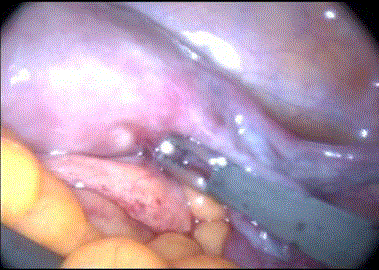
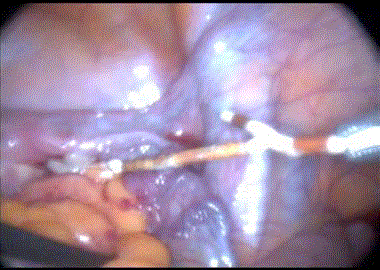
A 19-year-old, 1-year-old female patient was admitted to our outpatient clinic with the complaint of continuous systolic staining. The patient had a history of vaginal delivery six months ago. The patient had no history of systemic disease or operation. Approximately 1 month ago, a midwife in the health center had an Intrauterine Device (IUD) (copper t cu 365). Laboratory values; hemogram, C-reactive protein was normal, β-HCG was negative. On gynecological examination with speculum: IUD ropes were not observed and there were menstrual spotting. In vaginal ultrasonography, IUD refractory myometrium was observed. The IUD echo gave hyperechogenicity embedded in myometry. There were no IUD strings. There was minimal fluid in the Douglas cavity. Laparoscopy was decided due to suspicion of IUD perforating uterus. In laparoscopy, there was a tight connection between the uterus posterior and left uteroovarian and bowel mesos, and the right uterus perforated the uterus from the kornual region and the tip of the right istmo-tubal mesosalpinx was exposed to the abdomen (Figure 1). The tip was captured by laparoscopic holder and IUD was withdrawn (Figure 2). If the intestines were intact, the intestines were perforated and could be seen in the bowel cavity. After hemorrhage control, abdominal access sites (umbilicus and both pelvic) were closed with primary sutures. The patient was discharged with recommendations after 1 day.
Discussion
Today, IUD is frequently used as a method of birth control. It was found that it was 9.4% in
developed countries and 16.4% in underdeveloped countries [2].
They have frequent complications such as bleeding, pain, inflammatory disease, unwanted
pregnancy [3]. Uterine rupture is a rare but very important complication. It has been reported that
1,000 cases of rupture due to IUD are seen between 1,3-1,6 [1].
The clinician's inexperience with uterine rupture, breastfeeding and postpartum period, uterine
anomaly, adenomyosis have been associated with conditions [4-6]. Andersson et al. [4] in their
study, 50 consecutive patients with uterine rupture after IUD insertion were examined and IUD was
reported to be seen in 8 cases after doctor and 42 cases after midwife insertion. In another study,
70% of the rupture occurred in the first 6 months after delivery and therefore it was reported that it
was safer to wait for the patient to wear after 6 months [6]. In our patient, it was learned that IUD was inserted by the midwife at the 5th month after delivery.
Clinical diagnosis: Radiological, ultrasonographic and
laparoscopic methods are used. Clinically it is usually asymptomatic
if it does not cause any damage to adjacent organs. Sometimes it can
cause pain or bleeding. As a test, ultrasound and abdominal film
should be the first preferred methods. Vaginal Ultrasonography is a
non-invasive, inexpensive, safe method. Tomography and magnetic
resonance imaging can be used in undiagnosed cases. We performed
ultrasonography and found that IUD is moving through the cavity
and providing reflex in myometrium.
Endoscopic procedures or open surgery can be performed in the
treatment. First of all, laparoscopic procedures are necessary. This is
due to less invasive pain, less cosmetic results, lower hospitalization
and lower cost. In addition, the tissue trauma and adhesions are less
than open surgery.
However, it is possible to find different results of laparoscopy in
literature. These may include laparotomy, laparoscopy and colostomy
[7,8]. Demir et al. In their study, IUD was removed in 8 women and
no patient required laparotomy. There was no problem in the control
examinations of the patients [9]. Özgün et al. [10], in 10 patients, they
were removed laparoscopically and returned to laparotomy in only
2 patients. We used laparoscopic method for IUD removal and we
did not encounter any complications. On the 1st postoperative day,
we discharged the patient without any problems. We also did not
encounter any problems during their follow-up.
As a result, the most important factor for IUD insertion is the
correct patient selection. We also think it should be worn in expert
hands. Complications should be taken into consideration and every
patient with IUD should be checked at certain time intervals.
Figure 1
Figure 2
References
- Arslan A, Kanat-Pektas M, Yesilyurt H, Bilge U. Colon penetration by a copper intrauterine device: a case report with literature review. Arch Gynecol Obstet. 2009;279(3):395-7.
- Akpınar F, Özgür EN, Yılmaz S, Ustaoğlu O. Sigmoid colon migration of an intrauterine device. Case Rep Obstet Gynecol 2014;2014:207659.
- [No authors listed]. Mechanism of action, safety and efficacy of intrauterine devices. Report of a WHO Scientific Group. World Health Organ Tech Rep Ser 1987;753:1-91.
- Andersson K, Ryde-Blomqvist E, Lindell K, Odlind V, Milsom I. Perforations with intrauterine devices. Report from a Swedish survey. Contraception. 1998;57(4):251-5.
- Harrison-Woolrych M, Ashton J, Coulter D. Uterine perforation on intrauterine device insertion: is the incidence higher than previously reported? Contraception. 2003;67(1):53-6.
- Caliskan E, Oztürk N, Dilbaz BO, Dilbaz S. Analysis of risk factors associated with uterine perforation by intrauterine devices. Eur J Contracept Reprod Health Care. 2003;8(3):150-5.
- Gill RS, Mok D, Hudson M, Shi X, Birch DW, Karmali S. Laparoscopic removal of an intra-abdominal intrauterine device: case and systematic review. Contraception. 2012;85(1):15-8.
- Nceboz US, Ozçakir HT, Uyar Y, Cağlar H. Migration of an intrauterine contraceptive device to the sigmoid colon: a case report. Eur J Contracept Reprod Health Care. 2003;8(4):229-32.
- Demir SC, Cetin MT, Ucunsak IF, Atay Y, Toksoz L, Kadayifci O. Removal of intra-abdominal intrauterine device by laparoscopy. Eur J Contracept Reprod Health Care. 2002;7(1):20-3.
- Ozgun MT, Batukan C, Serin IS, Ozcelik B, Basbug M, Dolanbay M. Surgical management of intra-abdominal mislocated intrauterine devices. Contraception. 2007;75(2):96-100.


