Research Article
Grouping Patients after Surgery by Minimum Sum of Squares Clustering Method
Milorad Paunoviæ*
Clinical Center of Serbia, Serbia
*Corresponding author: Milorad Paunoviæ, Clinical Center of Serbia, Belgrade, Serbia
Published: 20 Sep, 2018
Cite this article as: Paunoviæ M. Grouping Patients after
Surgery by Minimum Sum of Squares
Clustering Method. World J Surg
Surgical Res. 2018; 1: 1061.
Abstract
In this paper for the first time the clustering method is applied in surgery post-operational
complications. We first collect data after surgery of 825 patients in hospital in Serbia, taking into
account 3 their attributes. Among 825 patients, 32 of them had the occurrence of dehiscence
laparotomy. We analyze the risk of taking surgery based on clustering patient in groups, taking into
account the presence of infection, diabetic and neoplastic disease. The minimum sum-of-squares
model is used and tested by k-means heuristic and Variable neighborhood search based method.
It appears that the method used is very important, i.e., VNS based heuristic provides significantly
better results. Some interesting conclusions are also derived.
Keywords: Clustering method, Surgery, Laparotomy, Grouping Patients
Introduction
Dehiscence of laparotomy is a sudden partial or complete opening or tearing wounds, or the formation of cracks in the surgical wound sewinged [1]. Complete wound disruption with evisceration of abdominal organs requires urgent re-intervention. It occurs most often during the first week after surgery. It occurs in 0.5% to 3% of operated patients [2]. Dehiscence of laparotomy is accompanied by high morbidity and mortality that ranges up to 40%. The process of wound healing is a highly complex and dynamic set of cellular, biochemical and immunological processes, which depends on several factors. Infection of the surgical wound is one of the most important risk factor for dehiscence of laparotomy. Gastrointestinal surgery, emergency surgery, prolonged surgical time, and are associated with an increased risk of surgical wound infection. Wound infection defined as purulent secretion from the wound contents, regardless of the bacteriological findings [3]. It occurs in up to 15% of treated patients [4-6]. Diabetes is characterized by atherosclerosis, microangiopathy, disorder of Hb dissociation and decreased chemotaxis and phagocytosis. Dehiscence of laparotomy is more common in patients with neoplastic diseases. Reasons are not entirely clear. It is assumed that the protein and calories lost in the tumor also has a premise that tumor cells secrete substances that interfere with wound healing [7]. The survey aims to determine the effect of the presence of infection, diabetes and malignant disease of the emergence of dehiscence of laparotomy.
Methods
Statistical tests
Research is organized by type of retrospective prospective studies that have analyzed the following
data as risk factors: The presence of infection, diabetes and neoplastic diseases of dehiscence of
laparotomy of 825 operated patients at the department of general surgery of Niš in 2016th and 2017th
year. Complications dehiscence of laparotomy was found in 32 patients. Statistical sample size is
determined by the statistical methodology to meet the basic principle of representativeness. The
normogram was used to determine the optimal sample. In this paper, results are presented in tables
and graphically. The statistical analysis using the methods of descriptive statistics (mean, standard
deviation), parametric tests (Student’s t-test) and nonparametric Chi square test. For statistical
analysis we used the software package SPSS 14.0 and the imaging table and a Microsoft Office Word
2003.
Minimum sum-of-squares clustering
One of mostly used criterion for clustering is Minimum Sum-of-Squares (MSS), where all
entities are placed in n-dimensional Euclidean space and their dissimilarities calculated as squared
distances in Rn. The number of clusters m is given in advance. The objective is to make groups
of entities such that the total sum of squared distances within each group or cluster is minimum.
It appears that minimizing the intra group distances is equivalent
to maximizing the square distances among entities from different
groups [8]. This property makes MSS most popular criterion since it
measures in the same time homogeneity and separation. Moreover,
MMS may be equivalently presented as the problem of minimizing
the square distances from each entity to its own cluster center or
centroid [8].
Since MMS problem is NP hard [8], there are many heuristics
already appeared in the literature? The most popular heuristic is so
called k-means method. It alternatively solves allocation of entities
to their closest centroid and finding the corresponding centroid of
each cluster. Although being very popular due to its simplicity, the
results obtained by k-means sometimes are very far from the global
optimum [8]. That is the reason why there are many heuristics that
are trying to improve precision of k-means algorithm. One among
them is J-means and Variable Neighborhood Search (VNS) based
heuristic [8].
In this paper we presented data of 825 patients in 3-dimensional
space. As mentioned earlier, those three attributes (or risk factors)
are: infection, diabetes, the presence of neoplastic diseases on the
occurrence of dehiscence of laparotomy. All three are considered
as binary variables. In the next section we will analyze the results
obtained by both k-means and VNS heuristics.
Table 1
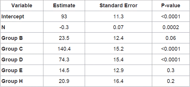
Table 1
Comparison of k-means and VNS heuristics in clustering n=825 patients into m groups.
Figure 1
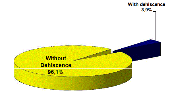
Figure 1
Number of dehiscence of the total number of respondents.
Figure 2
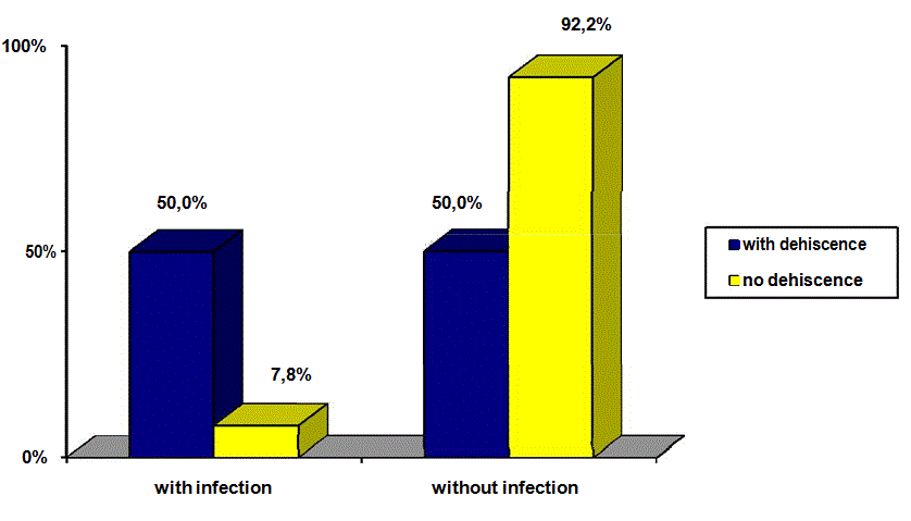
Figure 2
Impact of infection on the occurrence of dehiscence of laparotomy.
Results
Statistical tests
Dehiscence of laparotomy occurred in 3.9% of patients and 32
patients of the total 825 respondents (Figure 1). There is a statistically
significant relationship between dehiscence of laparotomy and
infections (χ2=62,024; p< 0.01). Infection was significantly more
prevalent in patients with dehiscence of laparotomy. Of 32 patients
with peritoneal them 16 or 50% had an infection and the 793 patients
without infection, dehiscence had all 62 of them, or 7.8% (Figure 2).
In the group of patients with dehiscence of laparotomy is more people
with diabetes than in the control group, but this was not statistically
significant (χ2=0.491; p>0.05). Patients with diabetes were 26 of them,
or 4.2% of the group of persons with dehiscence of laparotomy, and
596 patients with diabetes were in the group of patients without
dehiscence of laparotomy or 95.8%. In patients with dehiscence
without diabetes was 3.0% or 6 patients, and without dehiscence of
laparotomy and 197 patients without diabetes or 97.0% (Figure 3).
There is a statistically significant relationship between dehiscence
of laparotomy and neoplastic diseases (χ2=42,196; p< 0.01). Of
the 18 patients with neo plastic disease, 6 of them had dehiscence
of laparotomy or 33.3%, and 12 patients h ad no dehiscence of
laparotomy, or 66.7%. Without malignant disease were 26 patients
with dehiscence of laparotomy, or 3.2% and 781 patients without
dehiscence of laparotomy or 96.8% (Figure 4). Of the 32 patients
with dehiscence of laparotomy them 6 or 18.75% had a malignant
disease and the 793 patients without dehiscence of 12 or 1.51% had
a malignancy.
Clustering results
In Table 1 we report results obtained by two heuristics for
minimum sum-of-squares clustering: k-means and VNS. The first the
number of desired clusters are given. The second line gives the value
of the objective function, while in column 3 we report the number
of entities in each cluster obtained by k-means. The next 3 columns
report the same values given by VNS. It appears that both methods
keep 32 patients with dehiscence laparotomy in the same cluster. The
difference in results starts after m=5, where the total sum of squares
are 92.07 and 76.71 obtained by k-means and VNS respectively.
Moreover, VNS keeps the 32 patients in the same cluster up to m=8.
This means that not only the clustering model is important but also
the method used.
Some observations regarding results reported at Table 1 are:
1. Clustering models and methods may be successfully used
in medicine in general and more particularly in Surgery in parallel
with statistical tests;
2. Hypotheses may be automatically derived, e.g., the 32
patients with dehiscence of laparotomy are kept in the same group
with up to 8 clusters;
3. Results obtained by clustering techniques are more rich in
a sense that they provide more information to practitioners: relations
between clusters, introduction of many patient’s attributes in analysis,
etc;
4. The clustering method used may play a significant role in
understanding the final results, i.e., VNS based heuristic outperform
significantly k-means heuristic for number of clusters grater or equal
to 5.
Figure 3
Figure 3
Effect of diabetes on the occurrence of dehiscence of laparotomy.
Discussion
In this section we first discuss our results obtained by statistical
tests and then comment on their relations with clustering.
Despite major advances in the understanding of the process of
wound healing physiology, surgical techniques and the application
of modern technologies and materials in surgery, the percentage of
impaired healing laparotomy is still high. Dehiscence of laparotomy
occurs in approximately 3% of patients. In a retrospective study
by Rodriguez-Hermosa JI et al. [9] from Spain in 57 patients or
0.45% of the total 12,622 patients who had undergone laparotomy
occurred in dehiscence of laparotomy. The Cracow study Kenig J et
al. [10] and associates with dehiscence of laparotomy occurred in 56
patients or 2.9% of our patients. Our results show that dehiscence
of laparotomy was present in 3.9% of patients and 32 patients of the
total 825 respondents. Preoperative preparation is an important stage
in the treatment of surgical patients and the adequacy of preoperative
depends on result of the operation, the incidence of complications
and mortality of patients. It is necessary that all the general condition
of the patients preoperatively stabilized and carry a minimum of
anesthesia and surgical preoperative whenever the patient’s condition
allows [11]. Both studies confirm recent views that chronological
age of more than sixty years ago, in itself is not a contraindication
for extensive operations in abdominal surgery [7,12-14]. Far more
important are the parameters that determine the biological age of the
patient: The patient’s general condition and ability to care for oneself
(performance status), nutritional status (Seltzerov index), as well as
the risk of anesthesia estimated ASA score [12]. Infection is extremely
destructive effect on the wound healing process by increasing the
production of cytokines and proteases, which disrupt the synthesis
of fibroblasts, and the stability of the wound [15]. Our study confirms
this claim, because patients with the presence of infection, far more
frequent respiratory failure. 50% of patients with dehiscence of
laparotomy occurred of infection. In Germany, a study was done by
Fleischer GM and all, dehiscence of laparotomy occurs in 5% to 10%
of patients with infection [16]. In our study, the percentage impact
of infection on the occurrence of dehiscence much higher. In India’s
study from Rajindra Hospital in Patiala only 4 (8%) of our wound
dehiscence patients were diabetics. These patients were given insulin
[17]. Of all diabetics in our study does not receive any insulin therapy,
and because I have this complication less pronounced. In patients
with diabetes, dehiscence of laparotomy occurs more frequently but
it is not statistically significant (p>0.05). The five year prospective
observational study was performed 7,224 operations in 4,197 patients
in South Australia, 196 had diabetes patients (4.7%). The incidence of
2 patients with diabetes appeared [18] and do not differ from those
without dehiscence (p=90), which is concordant with our study. In
our study group of patients with dehiscence of laparotomy is more
people with diabetes than in the control group, but this was not
statistically significant (χ2=0.491; p>0.05). Patients with diabetes
were 26 of them, or 4.2% of the group of persons with dehiscence
of laparotomy, and 596 patients with diabetes were in the group of
patients without dehiscence of laparotomy or 95.8%. In patients with
dehiscence without diabetes was 3.0% or 6 patients, and without
dehiscence of laparotomy and 197 patients without diabetes or 97.0%.
The presence of malignant disease is accompanied by intense tumor
metabolism, malnutrition and disorders of absorption, all of which
can disrupt the normal wound healing process and therefore the more
frequent occurrence of dehiscence. Many drugs, anti-tumor and anticancer
drugs affect the various stages of wound healing, particularly
in cell division [14]. Statistically there is a strong association between
dehiscence of laparotomy and neoplastic diseases (χ2=42,196; p< 0.01).
Neoplastic diseases, in our material had 6 patients with dehiscence of
laparotomy, or 33.3%. A study worked in South Korea in 1987 to 2004
was included 8033 patients. Dehiscence of laparotomy occurred in
9.3% of treated patients with cancer [19]. Aksamija G et al. [20] who
worked on the study in Clinic for abdominal surgery in Sarajevo in
1998 to 2002 dehiscence after laparotomy surgery colon cancer from
439 patients were observed in 10 patients, or 2.27%.
Comparing the results with the results of international studies in this paper comes to the conclusion that our results are not worse than
the results of the world’s health task.
Figure 4
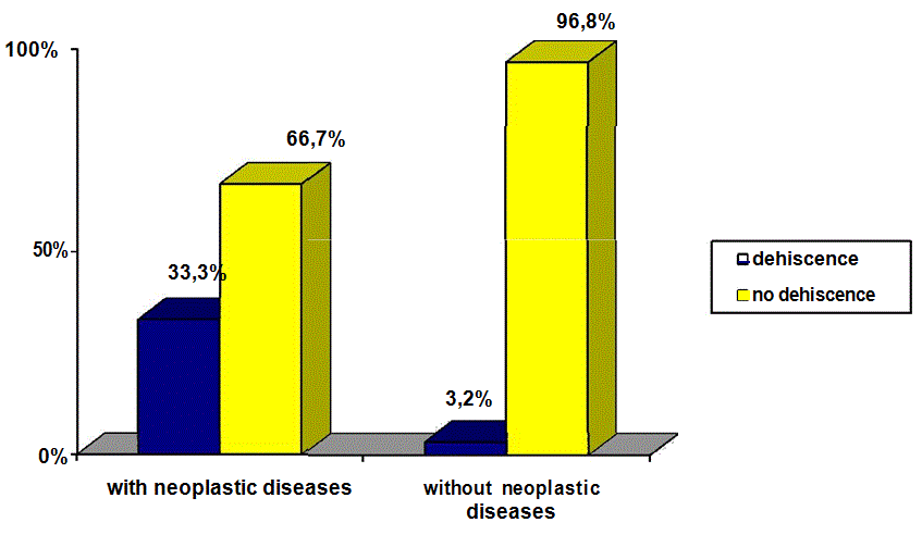
Figure 4
The presence of neoplastic diseases on the occurrence of
dehiscence of laparotomy.
Conclusion
Dehiscence of laparotomy occurs in less than 5% of patients.
In the presence of infection in patients with neoplastic diseases,
dehiscence of laparotomy is common. Dehiscence of laparotomy is
less common in people with diabetes. The analysis of these three risk
factors, the surgeon can identify patients at high risk and to take all
measures that prophylaxis his disposal.
In this paper we introduce for the first time a clustering technique
in analyzing risk factors on the occurrence of dehiscence laparotomy.
We show that minimum sum-of squares clustering model is well
suited for this purposes. Moreover, we show that some hypotheses
may be automatically derived, instead of assuming their validity
and then testing in usual statistical way. Future work may consist of
further implementation of other clustering paradigms in surgery, as
well as in medicine in general.
References
- Prpic Ivan, Surgery for pediatricians. School book. Zagreb; 2005;42-3.
- Radovanovic S. Abdominal hernia. Prosveta, Pozˇarevac, 1988;119-23.
- Brennan TG, Jones NAG, Gillou PJ. Lateral paramedian incision. Br J Surg. 1987;74(8):736-7.
- Hodgson N, Malthaner R, Ostbye T. The Search for an ideal method of abdominal fascial closure: A meta-analysis. Ann. Surg. 2000;231(3):436-42.
- Cleveland RD, Zitsch RP, Laws HL. Incisional closure in morbidly obese patients. Am Surg. 1989;55(1):61-4.
- Niggebrugge AH, Trimbos JB, Hermans J, Steup WH, Van De Velde CJ. Influence of abdominal-wound closure technique on complications after surgery: A randomized study. Lancet. 1999;353(9164):1563-7.
- Gerzic´ Z. Possible complications in digestive surgery. Institute for textbooks, Belgrade, 2000;625-9.
- Aloise D, Hansen P. On the complexity of minimum sum-of-squares clustering. GERAD. 2007;50:1-12.
- Rodriguez-Hermosa JI, Codina-Casadora, Ruiz B, Roig J, Girones J, Pujadas M, et al. Risk factors for acute abdominal wall dehiscence after laparotomy in adults. Cir. Esp. 2005;77(5):280-6.
- Kenig J, Richter P, Zurawska S, Lasek A, Zbierska K. Risk factors for wound dehiscence after laparotomy - clinical control trial. Pol Przegl Chir. 2012;84(11):565-73.
- Višnjic´ M, Surgery, Medical Faculty of Niš. DIGP ”PROSVETA” Niš, 2005;15-7.
- Zinner M: Maingot’s abdominal operations. Appleton Lange, London, 1997;548-80.
- Krivokapic´ Z, Prevention of colon anastomose relax. Science book, Beograd, 1990;39-47.
- Israelsson LA. Abdominal closure and incisional hernia. Eur. Surg. 2003;35:5-11.
- Jeremic´ M, Abdominal Surgery I, Medical Faculty, University of Niš. PELIKAN PRINT-Niš, 2009;23-8.
- Fleischer GM, Rennert A, Ruhmer M. Infected abdominal wall and burst abdomen. Chirurg. 2000;71(7):754-62.
- Ramnesh G, Sheerin S, Surinder S, Bar S. A Prospective Study of Predictors for Post Laparotomy Abdominal Wound Dehiscence. J Clin Diagn Res. 2014;8(1):80-3.
- Dixon AJ, Dixon MP, Dixon JB. Prospective study of skin surgery in patients with and without known diabetes. Dermatol Surg. 2009;35:1035-40.
- Oh SJ, Choi WB, Hyung WJ, Choi SH, Noh SH. Complications requiring reoperation after gastrectomy for gastric cancer: 17 years experience in a single institute. 2009;13(2):239-45.
- Aksamija G, Tanovic´ H, Sabanovic J, Muhovic´S. Early surgical complications after treatment of malignant diseases of the colon and rectum. Clinic for abdominal surgery, KCV Sarajevo, Med. Arh. 2003;57(3):23-4.