Research Article
Treatment of Fibroadenoma by Ultrasound-Guided Vacuum Assisted Breast Biopsy at Ho Chi Minh City Oncology Hospital
Phuong Viet The Tran1*, Cuc Hong Le2, Huong Thien Pham3 and Tu Hoang Pham4
1Department of Breast Surgery-Ho Chi Minh City Oncology Hospital, Vietnam
2Department of Radiology-Ho Chi Minh City Oncology Hospital, Vietnam
*Corresponding author: Phuong Viet The Tran, Department of Breast Surgery, Vinmec Hospital, 139/7A Phan Dang Luu Street, Ward 2, Phu nhuan District, Ho Chi Minh City, Vietnam
Published: 24 Aug, 2018
Cite this article as: Tran PVT, Le CH, Pham HT, Pham
TH. Treatment of Fibroadenoma by
Ultrasound-Guided Vacuum Assisted
Breast Biopsy at Ho Chi Minh City
Oncology Hospital. World J Surg
Surgical Res. 2018; 1: 1046.
Abstract
Introduction: VABB is effective for complete removal of benign lesion of the breast such as
fibroadenoma.
Method: breast lesions which are diagnosed fibroadenoma by ultrasound and less than 3 cm are
removed by ultrasound-guided VABB and local anesthesia.
Result: 20 patients with 39 lesions are removed with ultrasound guided VABB. All histology turns
out benign with 22 fibroadenomas. Mean age is 37.5 (11-54), mean size is 16.7 mm (9.5 mm to 30
mm). 8 cases with one tumor (40%), 6 cases with 2 tumors (30%), 5 cases with 3 tumors (25%) and
1 case with 4 tumors (5%). There is no severe complication, 5 cases with skin echymosis and 1 case
with hematoma. None of the patients feels pain.
Conclusion: VABB is an effective for diagnosis and treatment of benign breast lesion, including
fibroadenoma with small size.
Keywords: Fibroadenoma; Breast biopsy; Ultrasound-Guided
Introduction
Fibroadenoma is the most common benign tumor of the breast. Treatment of this type of tumor
is either follow up or removal. Follow up is often for small tumors, multiple tumors, young patients,
or patients who do not like surgery. Conventional treatment for tumor removal is open surgery.
This type of technique is relatively simple, with local anesthesia (if tumor is not big). However,
this technique has disadvantages such as leaving scars is an invasive method, and post-op care is
necessary.
Vacuum Assisted Breast Biopsy (VABB) has been performed worldwide since the 1990’. This
technique uses bigger needle (7G, 8G, 11G) and could biopsy or completely remove small benign
tumor (less than 3 cm). Therefore, this is a method for biopsy of breast lesions and also for treatment
of benign breast lesions. This technique has been approved by FDA (USA) and NICE (UK) for
complete removal of fibroadenoma.
In our knowledge, VABB has not been performed and reported in Vietnam until 2017. In this
article, we report our series of breast tumors diagnosed fibroadenomas by ultrasound and treated
with VABB at Ho Chi Minh City Oncology Hospital in 2017, which is the first reported series in
Vietnam.
Method
Patients who have breast tumors diagnosed fibroadenoma on ultrasound and less than 3 cm
are recruited. Patients will have consultation about options of follow up, open surgery or tumor
removal with VABB, pros and cons of each option.
When the patient decides to have VABB, her breasts are examined again with ultrasound by
the radiologist of the team. The character of the tumors, number and the size of the tumors are
recorded. Patients are also informed about the number of the tumor removed. If there are multiple
tumors, we decide to remove 2 to 3 tumors, those smaller than 10mm will be followed up. The
number of the removed tumor is also decided by the patient.
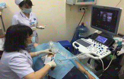
The procedure is performed at the Radiology department. Before
the procedure, breast ultrasound will be done to decide the entry
of the probe. Local anesthesia is with 20 ml of Lidocain 1% (with
Adrenalin) by 25G needle and then 18G needle. Lidocain is injected
behind and above the tumor. The purpose of Lidocain injection is
local anesthesia, homeostasis and to prepare the route for the probe.
The procedure is performed by radiologists or breast surgeons
who have been trained with VABB. A small orifice will be made by
an 11 blade to insert the probe. The size of the probe will be depended
on the size of the tumor and if the tumor is close to the skin or not.
The probe is inserted behind the tumor, at 6:00 position. When the
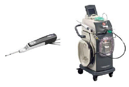
position of the probe is accurate, the Legacy machine (Mammotome-
USA) is activated and will cut and remove the tumor. The process of
removal of the tumor is visualized under ultrasound until the tumor
is removed completely and the probe is withdrawn afterward.
A nurse will compress the breast in 5 min for homeostasis and
the orifice is tapped. Patient stays at the hospital in 30 min and will be
instructed to remove the tap the day after. Patient will take pain killers
if painful but antibiotics are not prescribed. Patient comes back in 10
days for histology result, clinical and ultrasound examination.
Pain is evaluated on the scale of 1-10. Evaluation is taken before
the procedure, after the procedure and 10 days after the procedure.
Table 1
Figure 1
Figure 2
Result
There are 20 patients (39 tumors) diagnosed fibroadenoma on
ultrasound and treated with VABB. Mean age is 37.5 (11-54). Mean
size: 16.7 mm (9.5 mm to 30 mm). There are 8 patients with 1 tumor
(40%), 6 patients with 2 tumors (30%), 5 patients with 3 tumors
(25%), and one patient with 4 tumors (5%).
Pathology results of 39 tumors are 22 fibroadenomas, other
pathology results are borderline phyllodes tumor, fibrocystic
changes, and intraductal papilloma and granulloma mastitis (Table
1). Four patients diagnosed fibroadenoma turn out benign lesion (not
fibroadenoma), 16 cases have at least one fibroadenoma.
Complications are mild, including 5 cases with ecchymose of
the skin of the breast, one case with hematoma and needs puncture.
There are no infection, bleeding or chest wall injury. All the patients
do not have pain in the duration of the procedure, after the procedure
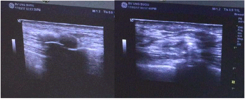
and 1 week after the procedure. Breast ultrasound shows that there is
no residue after the procedure and 10 days afterwards.
Discussion
Fibroadenoma is the most common tumor of the breast. For a
long time ago, treatment for this kind of tumor is follow up or opens
surgery. Follow up are for young patients, small tumor (<1 cm) and
multiple lumps. However, some patients worry about the tumor and
want to have the tumors removed for histology and VABB will give
the patients a good option because it is less painful, does not leave a
big scar and multiple lumps could be removed in one time.
VABB has been performing since 1995 and becoming an efficient
device for biopsy of breast lesions. VABB is useful for lesions small
than 5 mm which are not applicable for core biopsy. The probes
of VABB are bigger enough to provide large sample for histology.
Not only for biopsy, VABB has been approved by FDA and NICE
for treatment of benign breast lesions like fibroadenoma [1]. In
these situations, VABB provides patients an alternative of open
surgery without big scar. VABB could also remove multiple tumors
in one time. Povoski [2] report a case in which VABB removes 14
fibroadenoma of a 21 year old patient.
In the report of Karol et al. [3] with 196 cases diagnosed
fibroadenoma by ultrasound, only 157 cases (80.1%) are fibroadenoma
on histology, and 2 cases are invasive carcinoma. In the series of
Thurley et al. [4] with 134 cases diagnosed benign in ultrasound, only
81 (60.4%) are fibroadenoma on histology. In our series, 75% are
fibroadenoma pathologically. The remaining cases are benign except
one borderline phyllodes. In this case, patient rejects the option of
wide excision and decides to have followed up.
In our series, we do not use VABB for tumor bigger than 30 mm
(mean size is 16.7 mm). When the tumor is too big, the vacuum force
and the aperture of the probe could not completely remove the tumor.
In the series of Karol et al. [3], the mean size of the tumor is 13.53
mm. Two big tumors (50 mm and 60 mm) could not be removed
completely.
Like Lui [5], we use the probe 11 for lesions smaller than 1cm và
8 for lesions bigger than 1 cm. The choice of the size of the probe also
depends on the position of the tumor and skin, if the tumor is very
close to the skin, probe 8 could not be used to protect the skin.
Similar the series of Sperber et al. [6] with 52 cases, we do not
have any patient with pain in the procedure or afterwards.
VABB seldom has complication and often mild. Two patients of
Sperber et al. [6] have bleeding and resolve with compress. In 2477
cases of Lee et al [7], only 3 have bleeding and compression could
resolve the problem. Only 19% of patients of Thurley et al. [4] have
echymoses. We have 5 cases with mild ecchymoses and only one case
needs withdrawal for hematoma.
VABB has some disadvantages. In Vietnam, higher cost of VABB
compared to open surgery is an obstacle. This is a sophisticated
technique and need learning curve, radiologists and breast surgeons
need to master ultrasound guided intervention technique. Bigger
tumors is relative contraindicative of this procedure.
Figure 3
Figure 4
Discussion
VABB is an efficient method for diagnosis and treatment of breast benign lesions, including small fibroadenoma. This is a promising and potential option for treating of fibroadenoma in Vietnam.
References
- NICE. Image-guided vacuum-assiste excision biopsy of benign breast lesion. 2006.
- Povoski SP. The utilization of an ultrasound-guided 8-gauge vacuumassisted breast biopsy system as an innovative approach to accomplishing complete eradication of multiple bilateral breast fibroadenomas. World J Surg Oncol. 2007;5:124.
- Karol P, Dawid M, Piotr N, Beata A, Elizabeth G, Sonia F, et al. Vacuumassisted core-needle biopsy as a diagnostic and therapeutic method in lesions radiologically suspicious of breast fibroadenoma. Rep Pract Oncol Radiother. 2011;16(1):32-5.
- Thurley P, Evans A, Hamilton L, James J, Wilson R. Patient satisfaction and efficacy of vacuum assisted excision of fibroadenomas. Clin Radiol. 2009;(64):381-5.
- Lui CY, Lam HS. Review of Ultrasound-guided Vacuum-assisted Breast Biopsy: Techniques and Applications. J Med Ultrasound. 2010;18(1):1-10.
- Sperber F, Blank A, Metser U, Flusser G, Klausner JM, Lev-Chelouche D. Diagnosis and Treatment of Breast Fibroadenomas by Ultrasound-Guided Vacuum-Assisted Biopsy. Arch Surg. 2003;138(7):796-800.
- Lee SH, Kim EK, Kim MJ, Moon HJ, Yoon JH. Vacuum-assisted breast biopsy under ultrasonographic guidance: analysis of a 10-year experience. Ultrasonography. 2014;33(4):259-66.


