Case Report
Pancreatic Head Adenocarcinoma in Preadolescence
Kumar P1, Mandal S2 and Sarin YK1*
1Department of Pediatric Surgery, Maulana Azad Medical College, India
2Department of Pathology, Maulana Azad Medical College, India
*Corresponding author: Sarin YK, Department of Pediatric Surgery, Maulana Azad Medical College, New Delhi, Delhi, 110002, India
Published: 24 Aug, 2018
Cite this article as: Kumar P, Mandal S, Sarin YK.
Pancreatic Head Adenocarcinoma in
Preadolescence. World J Surg Surgical
Res. 2018; 1: 1041.
Abstract
Pancreatic cancers usually presents in older age group, with a median age of 66 years. Periampullary
tumors may arise from ampulla, distal common bile duct, pancreas or duodenum. We present
here a case of 12-year old girl with pancreatic head carcinoma.
Keywords: Pancreaticoduodenectomy; Pancreatic cancer; Whipple; Periampullary carcinoma
Introduction
Periampullary carcinomas are tumors arising within 2 cm diameter of ampulla of Vater and it comprises of carcinomas arising from ampulla, distal common bile duct, pancreas or duodenum. They share common presenting symptoms and therapeutic approaches. Here we present a case report of pancreatic head carcinoma presenting in a pre-adolescent girl and its management.
Case Presentation
A 12-year-old girl presented with complains of yellowish discoloration of eyes and skin for last
2 months. She gave history of on and off pain abdomen associated with fever, along with significant
weight loss. There was no history of clay colored stools. On clinical examination, she had pallor and
icterus. Per abdomen examination was within normal limits except for slight pain on deep palpation
in epigastrium. MRCP done elsewhere showed significantly dilated bi-lobar intra-hepatic biliary
radicals, Common Hepatic Duct (CHD) and Common Bile Duct (CBD) with abrupt narrowing of
distal CBD-likely CBD stricture. Multiple discrete and confluency nodular lesions involving both
hepatic lobes were present. Her hemoglobin was 8.4 g/dl and total bilirubin 5.8 g/dl at presentation.
Other liver function tests were normal with slight raise of SGOT and SGPT.
An ERCP was done which revealed ulceroproliferative growth at papilla and took multiple
biopsies which revealed moderately differentiated adenocarcinoma. Meanwhile, CECT abdomen
done at our center revealed a periampullary mass involving 2nd and 3rd parts of duodenum, head
of pancreas and distal CBD, with few ill-defined lesions in right lobe liver that could be metastases.
Hospital tumor board consultation was sought in view of suspected systemic metastasis; it
was decided to administer neo-adjuvant chemotherapy after decompression of biliary system. The
attempts at cannulation of CBD failed in view of bulky growth and distortion of anatomy of papilla.
A Percutaneous Transhepatic Biliary Drainage (PTBD) was successful with 8 Fr plastic stent, distal
end across ampulla. It helped in reduction of serum bilirubin from 11 g (raised meanwhile) to 2.9 g
and neo adjuvant chemotherapy was started, in form of gemcitabine. She received two cycles of neoadjuvant
chemotherapy and then radiological assessment was done using CT scan, which showed
resolution of liver lesions but static periampullary tumor without any large vessel involvement or
encasement.
She underwent Pylorus-Preserving Pancreaticoduodenectomy (PPPD) with gastrojejunostomy,
choledochojejunostomy and pancreaticojejunostomy. The presence of three polyps in duodenum
and jejunum was noted at surgery. Patient shifted to ICU in post-operative period and received
packed red cell volume and fresh frozen plasma transfusions. She developed ventilator associated
pneumonia and sepsis in post-operative period and succumbed on 5th postoperative day.
Histopathologic examination revealed it to be pancreatic head adenocarcinoma with R0 margins
and polyps were reported as tubular adenoma with no dysplastic changes. One lymph node was
positive.
Discussion
Periampullary carcinomas are increasingly recognized condition in view of increasing
awareness of symptoms, education level of patients and availability
of endoscopy and other radiological tools. It is a common name
given to tumors arising with in 2 cm of ampulla of Vater. Pancreatic
cancers usually present in older age group, with a median age of
66 years [1]. Periampullary carcinomas usually present with mass
effects, depending on organ of origin or secondary to metastatic
complications. There may be mechanical duodenal obstruction
causing gastric outlet blockage or compressing ampulla of Vater
causing jaundice, yellowish discoloration of body or hemorrhage
secondary to venous obstruction. Metastasis to liver or peritoneum
may cause ascites and liver cell failure. This child presented with
icterus and maintained liver function with increased Serum Bilirubin
(SB).
MRCP and sectional MR imaging are quoted to be useful in
determining the origins of periampullary carcinomas [2]. There
are various signs which help in differentiating organ of origin, like
pancreatic cancer usually demonstrate dilatation of side branches of
pancreatic duct and enhance poorly on gadolinium enhanced images
and ampullary carcinomas manifests as a small mass, periductal
thickening, or bulging of the duodenal papilla. These findings could
not be appreciated in our patient in view of poor quality films and nonavailability
of 3D reconstructed images. CECT and CT angiography
with 3D reconstruction has been also quoted in literature to guide
resectability of cancer [3]. Grade 4 lesions (total encasement of either
the superior mesenteric vein or artery) were considered unresectable.
Resectability rates decreases as the grade of lesion increases. Our
patient had grade 2 lesion on CT scan, with resectability rate of 50%
as quoted in literature [3].
Endoscopy surveillance and ablative therapy has role in
management of periampullary lesions especially in adenomas [4],
although eradication of ampullary lesion may need multiple settings.
Literature also quotes of adenoma- carcinoma sequence in duodenum
in familial adenomatous polyposis syndrome in adults [5], though no
recognized genetic syndromes associated with pancreatic carcinoma
in children or adolescents [6-9]. Although our patient had three
polyps in duodenum and jejunum, which histologically came out
to be tubular adenoma with no dysplastic changes, so any genetic
correlation could not be sought.
Preoperative biliary drainage was introduced to decrease
bilirubin load, help in symptomatic palliation in metastatic disease
or render patient fit for chemotherapy and surgery. But two metaanalyses
showed that patients receiving pre-operative biliary drainage
have more post-operative complications [10,11]. Success of biliary
drainage is documented to be decrease of >50% of SB within 2 weeks
and proceeding to surgery in next 4 to 6 weeks [12], which could not
be achieved in our patient in view of twice shunt blockage.
Preoperative platelet counts are also identified in literature as
prognostic factor in periampullary carcinoma. Low platelet counts
associated with inferior outcome results, mechanisms unclear, may
be general factors or platelet specific factors [13].
The agents of metastatic pancreatic cancer include 5-FU,
streptozotocin, mitomycin C, gemcitabine and doxorubicin,
associated with response rates of 7% to 36% [14-16]. Our patient
received 2 cycles of gemcitabine chemotherapy. Response may be
quoted as we could do R0 resection and did not find any encasement
of great vessels [CT showed grade 2 lesions].
Our patient underwent pylorus preserving
pancreaticoduodenectomy as there was no gross infiltration of D1
or distal stomach. Literature states that pylorus preservation does
neither compromise long-term survival, nor increase any operative
risks [17].
In one of the largest single-institution experience with
Pancreaticoduodenectomy [PD] for pancreatic cancer, median age
was 66 years, median tumor diameter was 3 cm, 42% of PD specimens
had positive margins, and 78% had positive Lymph Nodes (LN), with
median survival of 18 months. They quoted peri operative morbidity
of 38% and decrease in mortality rates of 38% in 1970s to 1% in 2000s
[1]. Factors having impact on survival were tumor diameter, LN
status, margins and histology grade. Pancreatic head adenocarcinoma
has poor prognosis as compared to non-pancreatic peri ampullary
carcinomas.
In a large UK experience of adenocarcinoma of the head of the
pancreas and periampullary region, age <60 years, tumor of head
of pancreas, positive LNs, R1 resection, poor differentiating tumor,
portal vein invasion were studies to be independent factors for
decreased survival. Mortality in study was 4.8%, with median survival
of 13.4 months in pancreatic head, 35.5 months in ampullary and
16 months in distal bile duct cancers [18]. Our patient had three
independent negative factors as per this study.
Simultaneous resection of peri-ampullary or pancreatic cancer,
with synchronous liver metastasis has been attempted but it has
proven to be associated with increase morbidity and mortality and
does not increase long term survival [19]. Our patient did not have
any gross lesions on liver surface and no liver procedure was done for
suspected metastasis lesion as per CT scan.
Figure 1
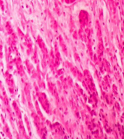
Figure 1
HPE showing duodenal mucosa along with tumor present in small
nests and sheets.
Figure 2
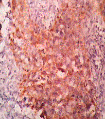
Figure 2
The nests and clusters of tumor cells with high NC ratio, scant
cytoplasm and prominent nucleoli.
Figure 3
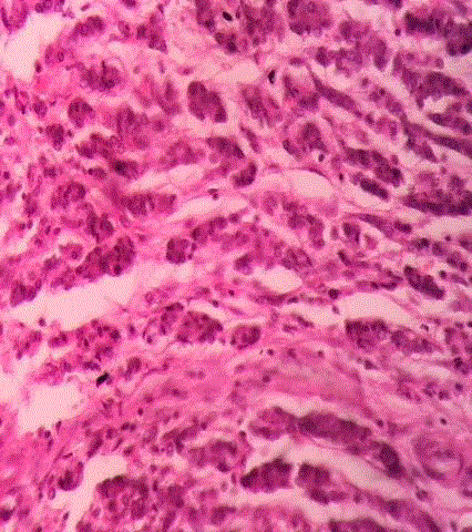
Figure 3
The nests and clusters of tumor cells with high NC ratio, scant
cytoplasm and prominent nucleoli.
Figure 4
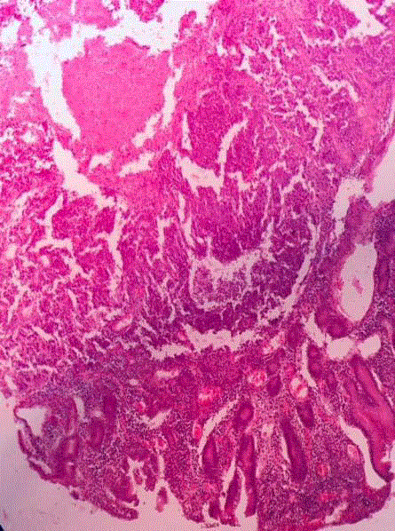
Figure 4
Tumor cells showing expression of cytokeratin on IHC.
References
- Winter JM, Cameron JL, Campbell KA, Arnold MA, Chang DC, Coleman J, et al. 1423 pancreaticoduodenectomies for pancreatic cancer: A singleinstitution experience. J Gastrointest Surg. 2006;10(9):1199-211.
- Kim JH, Kim MJ, Chung JJ, Lee WJ, Yoo HS, Lee JT. Differential diagnosis of periampullary carcinomas at MR imaging. Radiographics. 2002;22(6):1335-52.
- Saldinger PF, Reilly M, Reynolds K, Raptopoulos V, Chuttani R, Steer ML, et al. Is CT angiography sufficient for prediction of resectability of periampullary neoplasms? J Gastrointest Surg. 2000;4(3):233-9.
- Norton ID, Geller A, Petersen BT, Sorbi D, Gostout CJ. Endoscopic surveillance and ablative therapy for periampullary adenomas. Amer J Gastroenterol. 2001; 96(1):101-6.
- Spigelman AD, Talbot IC, Penna C, Nugent KP, Phillips RK, Costello C, et al. Evidence for adenoma-carcinoma sequence in the duodenum of patients with familial adenomatous polyposis. The Leeds Castle Polyposis Group (Upper Gastrointestinal Committee). J Clin Pathol. 1994;47(8):709- 10.
- Hayman W, Neerlaub RC, Johnson TS. Pancreatic carcinoma in childhood: Report and review. J Pediatr. 1974;65:1711.
- Lack EE, Cassady JR, Levey R, Vawter GF. Tumors of the exocrine pancreas in children and adolescents. A clinical and pathologic study of eight cases. Amer J Surg Pathol. 1983;7(4):319-27.
- Grosfeld JL, Vane DW, Rescorla FJ, McGuire W, West KW. Pancreatic tumors in childhood: analysis of 13 cases. J Pediatr Surg. 1990;25(10):1057- 62.
- Warshaw AL, Castillo CF. Pancreatic carcinoma. N Engl J Med. 1992;326(7):455-65.
- Wang Q, Gurusamy KS, Lin H, Xie X, Wang C. Preoperative biliary drainage for obstructive jaundice. Cochrane Database Syst Rev. 2008;(3):CD005444.
- Sewnath ME, Karsten TM, Prins MH, Rauws EJ, Obertop H, Gouma DJ. A meta-analysis on the efficacy of preoperative biliary drainage for tumors causing obstructive jaundice. Ann Surg. 2002;236(1):17-27.
- van der Gaag NA, Rauws EA, van Eijck CH, Bruno MJ, van der Harst E, Kubben FJ, et al. Preoperative biliary drainage for cancer of the head of the pancreas. N Engl J Med. 2010;362(2):129-37.
- Schwarz RE, Keny H. Preoperative platelet count predicts survival after resection of periampullary adenocarcinoma. Hepatogastroenterology. 2001;48(41):1493-8.
- Evans DB, Abbruzzese JL, Rich TA. Cancer of the pancreas. In: DeVita VT Jr, Hellman S, Rosenberg SA, editors. Cancer: Principles and Practice of Oncology. 5th ed. Philadelphia: JB Lippincott; 1997. p. 1054.
- Moore M. Activity of gemcitabine in patients with advanced pancreatic carcinoma: a review. Cancer. 1996;78(3):633-8.
- Vossen S, Goretzki PE, Goebel U, Willnow U. Therapeutic management of rare malignant pancreatic tumors in children. World J Surg. 1998;22(8):879-82.
- Grace PA, Pitt HA, Tompkins RK, Denbesten L, Longmire WP. Decreased morbidity and mortality after pancreatoduodenectomy. Am J Surg. 1986;151(1):141-9.
- Jarufe NP, Coldham C, Mayer AD, Mirza DF, Buckels JA, Bramhall SR. Favorable prognostic factors in a large UK experience of adenocarcinoma of the head of the pancreas and periampullary region. Dig Surg. 2004;21(3):202-9.
- Gleisner AL, Assumpcao L, Cameron JL, Wolfgang CL, Choti MA, Herman JM, et al. Is resection of periampullary or pancreatic adenocarcinoma with synchronous hepatic metastasis justified? Cancer. 2007;110(11):2484-92.