Case Report
A Case of Acute Suprascrotal Vasitis
Matthew Megson*, Adam Jones and Sid Singh
Department of Urology, George Eliot Hospital, UK
*Corresponding author: Matthew Megson, Department of Urology, George Eliot Hospital, College Street, Nuneaton, CV10 7DJ, UK
Published: 24 Aug, 2018
Cite this article as: Megson M, Jones A, Singh S. A Case
of Acute Suprascrotal Vasitis. World J
Surg Surgical Res. 2018; 1: 1037.
Abstract
A 35-year-old man presented with an acute, painful lower left quadrant and groin mass with signs
of sepsis. On examination, it was difficult to tell whether this was a strangulated hernia or collection.
Biochemical investigations revealed raised inflammatory markers and radiological investigations
showed a rare inflammatory condition, acute suprascrotal vasitis, which is mistaken for various
other 'surgical' groin masses.
This case report summarizes the importance of realizing this differential diagnosis for an acute groin
masses and how imaging can prevent unnecessary surgery.
Case Presentation
This 35 year old gentleman presented to A & E on the 27th of April, 2017 with a 5/7 day history
of left lower quadrant pain which was constant in nature, increasing in intensity, and worsened by
movement. There was no association with eating or opening bowels, he had no lower urinary tract
symptoms, though he did show signs of sepsis. He had experienced rectal bleeding in the evening
for the preceding week, and night sweats. He had a recent diagnosis of IBD (Inflammatory Bowel
Disease) and IDDM (Insulin Dependent Diabetes Mellitus). He developed a swelling in his left
groin, which was not reducible and had mild skin changes overlaying this. On examination, there
were no scrotal signs of epididymitis, he was tender left lower quadrant and over his left inguinal
canal. An USS (Ultrasound Scan) showed a no hernia, but inflammation within his inguinal canal.
He was put on ciprofloxacin to cover possible epididymitis. Despite this he did not improve over
the next week, so a CT abdomen/pelvis was done (Figure 1), showing a collection and inflamed vas
from inguinal canal to seminal vesicles.
He was changed to IV Meropenem for 72 hours, then stepped down to a higher dose of oral
ciprofloxacin. His collection was drained using US guided aspiration. This gentleman was discharged
home, and had his US guided aspiration of his collection as an outpatient procedure (Figure 2).
Investigations
USS-both testes and epididymi are symmetrical in size shape and echogenicity as well as color
Doppler imaging. There is a small amount of left sided hydrocele and varicocele. There is diffuse
thickening and enlargement of the left inguinal canal with mild increased vascularity within it. I
suspect this represents an inflamed spermatic cord.
CT (Figure 1)-normal liver, spleen, pancreas and both adrenals and both kidneys are normal
calibre of the aorta, no paraaortic lymphadenopathy. Faecal residue is seen round the colon. The
rest of the abdomen and pelvis is unremarkable. In the left groin region extending to the scrotum
there is a localized fluid collection seen which measures approximately 5.9 cm × 3.2 cm in size .The
exact nature of this collection is not clear? Cyst around the spermatic cord/? Localized abscess,
USS (Figure 2)-there is a dense-fluid-collection measures approx 1.7 cm × 2.8 cm × 4.8 cm situated
medially and dorsally to the spermatic cord in the upper inguinal canal. There are few tiny septation
inside this collection.
Differential Diagnosis
Originally he was diagnosed with acute exacerbation of his IBD, as he was admitted with PR bleeding, high white cell count, and left lower quadrant pain. Due to the swelling in his groin he was then diagnosed to have a hernia which was excluded on the USS. Due to the swelling if his groin and the suspicion of the sonographer that he had an inflamed spermatic cord within his inguinal canal he was diagnosed with epididymo-orchitis, though due to the lack of scrotal signs this was ruled out. The CT scan was done to investigate if there was an intra-abdominal cause for his inguinal inflammation, though this proved acute suprascrotal vasitis.
Figure 1
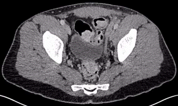
Figure 1
This is a CT axial view through this patients’ pelvis. This shows the
inflamed left vas originating from the inguinal canal.
Figure 2
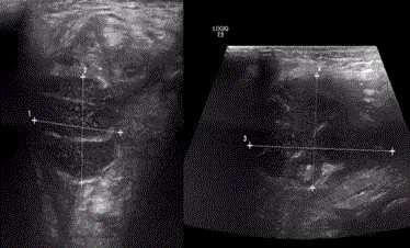
Figure 2
This is an USS of the collection in this patients’ left groin.
Discussion
The condition was first described in 1795 by Benjamin Bell [1].
It has often been confused with epididymitis, orchitis, testicular
torsion, or inguinal hernia, with the correct diagnosis usually being
made at the time of surgery [2], this surgery being unnecessary. On
USS acute vasitis usually presents with infection combined with acute
epididymitis, and it usually appears as a heterogeneously hypoechoic
lesion in the scrotal segment, suprascrotal segment, or both [3]. Most
of the literature denotes patients who have scrotal signs or have
epididymitis as well as vasitis [4-9]. Whereas our patient had very
minimal scrotal signs, which made the diagnosis more challenging
to come to.
The Pathogens are similar to those of epididymitis and are usually
not isolated in the urine. For those patients less than age 40 years
of age it could be an STI pathogen (Chlamydia, N gonorrhea) [10],
or a UTI pathogen (Escheria coli, Haemophilus influenzae) [11]. For
those patients over the age of 40 it is more likely to be UTI pathogens.
Rare pathogens also described in case reports include Mycobacterium
tuberculosis and Schistosoma haematobium [12,13].
In summary, this is a case of a rare condition which is often
misdiagnosed. There is often confusion over the diagnosis which may
result in unnecessary surgery. However, this is a condition which can
be treated medically with antibiotics and therefore clarification of the
diagnosis is essential. This is a diagnosis which should be thought of
as a possibility for those patients presenting with an acute groin mass.
Learning Points
1. There are many differential diagnoses for an acute groin
mass, some of which do need urgent surgical intervention, others it’s
inappropriate to operate on.
2. If the patient does not improving during treatment for your
working diagnosis then there is a need to re-evaluate the diagnosis.
3. This is likely an under reported diagnosis and therefore it
should form part of your differential diagnosis of an acute groin mass.
4. A good sonographer/radiologist may be able to distinguish
this on USS, however if not then a CT may be warranted.
References
- Bell B. A treatise on gonorrhea virulenta and lues venerea. Philadelphia: Printed for Robert Campbell, bookseller; 1795.
- Eddy K. Acute Infectious Vasitis: Potentially Under-reported Condition Mimicking Inguinal Hernia. DAL. 2015.
- Yang DM, Kim HC, Lee HL, Lim JW, Kim GY. Sonographic findings of acute vasitis. J Ultrasound Med. 2010;29(12):1711-5.
- Bissada NK, Redman JF, Finkbeiner AE. Unusual inguinal mass secondary to vasitis. Urology. 1976;8(5):488-9.
- Maitra AK. Odd inguinal swelling. Lancet. 1970;1(7636):45.
- Ryan SP, Harte PJ. Suppurative inflammation of vas deferens: an unusual groin mass. Urology. 1988;31(3):245-6.
- Wolbarst AL. The vas deferens, a generally unrecognized clinical entity in urogenital disease. J Urol. 1933;29(4):405-12.
- Wilson SR, Katz DS. Computed tomography demonstration of epididymitis with extension to vas deferens. Urology. 2006;68(6):1339-40.
- You SH, Sung DJ, Han NY, Park BJ, Kim MJ. Emphysematous vasitis misdiagnosed as strangulated inguinal hernia. J Emerg Med. 2014;47(1):e15-7.
- Dylewksi J, Sygal V. Infectious vasitis caused by Chlamydia trachomatis. Infec Dis ClinPract. 2014;22(3):e16-7.
- Chan PT, Schlegel PN. Inflammatory conditions of the male excurrent ductal system. Part II. J Androl. 2002;23(4):461-9.
- Yang DM, Kim HC, Kim SW, Lee HL, Min GE, Lim SJ. Sonographic findings of tuberculous vasitis. J Ultrasound Med. 2014;33(5):913-6.
- Durand F, Brion JP, Terrier N, Pinel C, Pelloux H. Funiculitis due to Schistosoma haematobium: uncommon diagnosis using parasitologic analysis of semen. Am J Trop Med Hyg. 2004;70(1):46-47.