Short Communication
A Rare Clinical Case of Multivessel Stenosis and Lesions in Patient with Takayasu's Disease
Kaliaev AO1, Bakhmetev AS2, Bakhmeteva MS3 and Malikova Marina4*
1Department of Radiology, Boston Medical Center, USA
2Department of Surgery, Saratov State Medical University named after V. I. Razumovsky, Russia
3Department of Surgery, Avesta Central Hospital, Russia
4Department of Surgery, Boston University, USA
*Corresponding author: Malikova Marina, Department of Surgery, Boston University, Boston Medical Center, Boston, Massachusetts, 02181, USA
Published: 06 Aug, 2018
Cite this article as: Kaliaev AO, Bakhmetev AS,
Bakhmeteva MS, Marina M. A Rare
Clinical Case of Multivessel Stenosis
and Lesions in Patient with Takayasu's
Disease. World J Surg Surgical Res.
2018; 1: 1033.
Abstract
Takayasu's disease is a systemic, inflammatory autoimmune disease. This disease is more often
affecting young or middle-age women of an Asian descent. It predominantly affects the aorta and its
branches, as well as the pulmonary arteries.
Keywords: Takayasu's disease; Multivessel lesions; Vascular ultrasound diagnostics
Introduction
Takayasu's disease, also known as Takayasu arteritis, is a form of large vessel granulomatous
vasculitis with massive intimal fibrosis and vascular narrowing. This disease is more often affecting
young or middle-aged women of an Asian descent. It predominantly affects the aorta and its
branches, as well as the pulmonary arteries. In terms of prevalence, females are about 8 to 9 times
more likely to be affected than males [1].
Takayasu's disease is similar to other forms of vasculitis, including giant cell arteritis which
typically affects older individuals [1,2]. Due to obstruction of the main branches of the aorta,
including the left common carotid artery, the brachiocephalic artery, and the left subclavian artery,
Takayasu's arteritis can present as pulseless upper extremities with weak or absent pulses on the
physical examination, which may be why it is also commonly referred to as the "pulseless disease”
[3].
Although the cause of Takayasu arteritis is unknown, the condition is characterized by segmental
and patchy granulomatous inflammation of the aorta and its major derivative branches. This
inflammation leads to arterial stenosis, thrombosis, and aneurysms [3]. There is irregular fibrosis of
the blood vessels due to chronic vasculitis, sometimes leading to massive fibrosis of the inner section
of the blood vessels (intima fibrosis) [3].
Clinical features of this disease can be detected on physical vascular examination of lower
extremities by assessing pulses and confirmed by ultrasonography. Diagnosis is based on the
demonstration of vascular lesions in large and middle-sized vessels on angiography, CT scan,
Magnetic Resonance Angiography (MRA). Contrast angiography has
been the gold standard. However, angiography provides information
on vessel anatomy and patency but does not provide information on
the degree of inflammation in the wall [2,3].
Ultrasonography remains a primary imaging modality to diagnose
vascular diseases, including Takayasu’s arteritis. It is cost efficient and
minimally invasive, since it doesn’t require use of contrast agents,
and/or anesthesia related to intervention [2,3]. The earliest detectable
on ultrasound lesion is a local narrowing or irregularity of the lumen.
This may develop into stenosis and occlusion. The characteristic
finding is the presence of "skip lesions," where stenosis or aneurysms
alternate with normal vessels. If such lesions are detected, further
evaluation on CT scan or MRA is recommended [2,3].
Figure 1
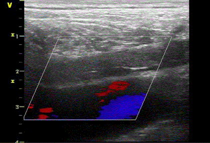
Figure 1
Occlusion of 50 mm with homogenous masses of medium echogenicity, which firmly adhered to the
vessel wall, detected in the right common iliac artery by ultrasonography.
Figure 2
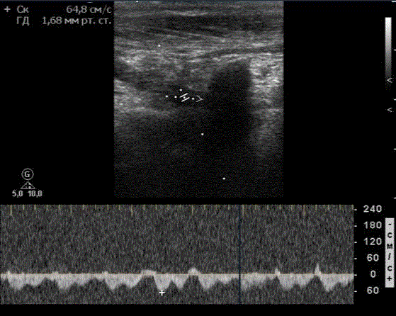
Figure 2
Steal syndrome was diagnosed based on retrograde blood flow in
vertebrate artery (shown by white arrowhead) and detected by ultrasound at
60 cm/second.
Figure 3
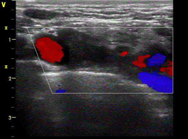
Figure 3
Occlusion of the first segment of subclavian artery as detected by
ultrasonography.
Purpose
To analyze and describe a rare clinical case of multivessel stenosis and lesions in Takayasu's disease of a 35 years old man.
Material and Methods
The patient was examined by utilizing stationary device Philips HD 11 expert class with phased sectoral sensor at 2 MHz to 4 MHz frequency at the Ultrasonic and Functional Diagnostics Department of the Mirotvortseva hospital, which is affiliated with Saratov State Medical University (SSMU).
Figure 4
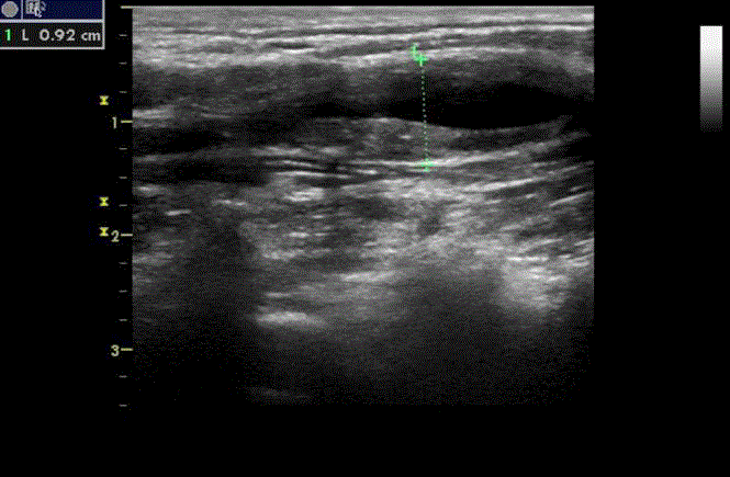
Figure 4
Bifurcation of common carotid artery. A diffused thickening of the
right common carotid artery walls, which was circular in shape (shown in
dotted line) as detected by ultrasound. The maximum stenosis detected by
ultrasonography was up to 60%.
Results
The 35-year-old man, who lives in the Saratov region, was referred
by a general surgeon to the Mirotvortseva hospital. The patient had
complaints of pain in the right leg shin, which appeared after walking
about 100 m. The onset of the disease was 3 months prior according
to the patient, when discomfort in the lower limb arose after walking
on average 450 m. The patient had asthenic physique, without bad
habits and chronic diseases. On physical examination there was no
pulsation on the femoral, popliteal and tibial arteries on the right side,
and only weak pulsations on the right radial artery was detected.
The triplex ultrasound scanning showed a 50 mm occlusion of
the right common iliac artery (Figure 1). There were homogenous
masses of medium echogenicity, which firmly adhered to the vessel
wall. The differentiation into layers was lost in the vessel (Figure
1). There was a collateral blood flow detected and thickness of the
intima-media complex was measured at no more than 0.65 mm in
distal levels of right limb’s arteries. As for the left limb, there were no
pathological changes seen on ultrasound and physical examination.
When we checked the brachiocephalic arteries, we found vertebral
subclavian steal syndrome (as shown on Figure 2) with occlusion
of the first segment of the subclavian artery (Figure 3), and diffused
thickening of the walls of the right common carotid artery, which
was circular in shape (Figure 4). The maximum stenosis detected
was up to 60% in the bifurcation of carotid artery (Figure 4). There
were no pathological changes found in the left branches of the aortic
arch, and the intima-media complex was measured at up to 0.65 mm
(not shown). The abdominal aorta and visceral branches were also
examined. The walls were not thickened; the blood flow was without
changes. The celiac trunk was not affected (not shown). According
to clinical and laboratory tests (e.g., normal coagulation and lipid
spectrum, insignificant increasing of the blood sedimentation rate and
C-reactive protein), relatively young age (35 years old), complaints of
intermittent claudication, weak pulsation on the radial artery, and the
presence of close relatives with Asian heritage which associated with
genetic predisposition, the patient’s diagnosis was determined to be
the Takayasu’s disease.
Conclusion
When identifying a non-atherosclerotic profile in young patients with the symptoms of intermittent claudication with occlusive lesions in the lower limbs, we recommend performing an ultrasound examination of brachiocephalic arteries for excluding the multivessel lesions involvement, which can be a sign of rarer incidence of an advanced and dispersed in different parts of the body Takayasu's disease.
References
- American College of Physicians (ACP). Systemic Vasculitis. Medical Knowledge Self-Assessment Program (MKSAP-15): Rheumatology. ACP. 2009;65-7.
- Johnston SL, Lock RJ, Gompels MM. Takayasu arteritis: A review. J Clin Pathol. 2002;55(7):481-6.
- Ishikawa K, Maetani S. Long-term outcome for 120 Japanese patients with Takayasu's disease. Clinical and statistical analyses of related prognostic factors. Circulation. 1994;90(4):1855-60.