Case Report
A Rare Tumor in the Colon: A Case of Pleomorphic Sarcoma
Akdeniz N, Urakci Z, Kaplan MA* and Isikdogan A
Department of Medical Oncology, Dicle University, Turkey
*Corresponding author: Kaplan MA, Department of Medical Oncology, Dicle University, Diyarbakir, 21280, Turkey
Published: 13 Jul, 2018
Cite this article as: Akdeniz N, Urakci Z, Kaplan MA, Isikdogan A. A Rare Tumor in the Colon: A Case of Pleomorphic Sarcoma. World J Surg Surgical Res. 2018; 1: 1024.
Abstract
Colonic primary mesenchymal tumors are very rare. Although they resemble epithelial tumors both clinically and macroscopically, they can be distinguished by their pathological features. They are treated with adjuvant chemotherapy and/or radiotherapy, depending on risk factors after resection. Rare cases of pleomorphic sarcomas should be considered in the differential diagnosis of patients presenting with colon cancer.
Keywords: Colon; Mesenchymal tumor; Pleomorphic sarcoma
Introduction
Ninety-five percent of colon cancers are of epithelial origin and develop on the basis of adenomatous polyps. In contrast, mesenchymal tumors of the colon are quite rare and form a heterogeneous group. Primary mesenchymal tumors of the gastrointestinal tract constitute only 0.1% to 3% of all gastrointestinal system cancers. Of the mesenchymal tumors, leiomyosarcoma is often seen in the colon while liposarcoma, pleomorphic sarcoma, synovial sarcoma, and desmoplastic small round cell sarcoma may also rarely be seen [1,2]. We report a case of pleomorphic sarcoma, a rare primary mesenchymal tumor of the colon.
Case Presentation
A 74-year-old male patient was consulted for abdominal pain and intermittent rectal bleeding for the past 4 months, and constipation for the past week. The patient's past history included hypertension, thyroidectomy, and coronary artery disease. His family history was not remarkable. On physical examination, his conjunctivae were pale, and he had diffuse abdominal tenderness. Laboratory tests showed an increase in C-reactive protein 6.68 mg/dL (reference: 0 to 0.5 mg /dL) and lactat dehidrogenaza 290 U/L (reference: 125 U/L to 220 U/L). His hemoglobin level was 10.4 gr/dL and hematocrit level was 32%. Tumor markers were within normal limits. An abdominal and pelvic computed tomography revealed an increased wall thickness of an approximately 10 cm segment of the sigmoid colon as well as a mass lesion of approximately 51 mm × 33 mm extending from the adjacent rectum on the right, permeating the mesenteric fascia, and extending to the adjacent muscle group. Similar mass lesions were observed on the right iliopsoas, at the site of the cecum, muscles adjacent to the cecum, right kidney’s posterior part, and the retroperitoneal area (Figure 1). The patient underwent colonoscopy, which revealed a massive mass with ulceration on the spot and near the lumen at 17th cm (Figure 2). As the clinical and imaging studies indicated obstruction, the patient was urgently taken to operation. A postoperative histopathological examination of the excised masses yielded a pleomorphic sarcoma as the primary diagnosis (Figure 3). The tumor was multiple; with a maximum diameter of approximately 7.5 cm. Necrosis was of grade 2 and less than 50%. Immunohistochemical study results were as follows; vimentin positive; CD68, CDX2 focal positive; SMA, S-100, CD117, desmin, CD20, LCA, CD34, CD31 negative; Ki-67 proliferation index was found to be 15% (Figure 4). The surgical margin was negative and metastases were detected in 3 out of 22 dissected lymph nodes. Pelvic computed tomography showed postoperative necrotic mass lesions in the posterior and medial pelvic regions, right iliopsoas, and right renal posterior and right pararenal fascias. In the rectosigmoid localization there were mass lesions passing the bladder posteriorly and mesorectal fascia and extending to the adjacent muscle group. The patient was considered to have metastatic disease and palliative chemotherapy was started. AIM treatment consisting of Adriamycin 60 mg/m², ifosfamide 2,500 mg/m², mesna 2,500 mg/m² was administered for 3 consecutive days and every 21 days thereafter. The patient was hospitalized upon the diagnosis of ileus while the treatment cycles continued. Perforation and peritonitis were detected in the imaging and the patient died after 6 months.
Figure 1A and B
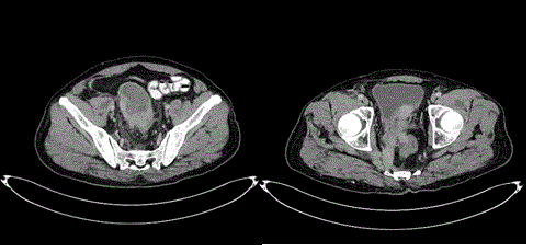
Figure 1A and B
Axial CT image; random massive thickness increase in sigmoid colon-the mass lesion extends to the bladder, to the pelvic region, and left-sided muscle groups.
Figure 2
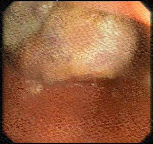
Figure 2
Colonoscopy shows an intraluminal giant mass of the left colon.
Figure 3
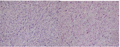
Figure 3
Eosinophilic cytoplasmic fusiform cells are seen in histopathological examination (Hematoxylin-eosin staining × 2oo).
Figure 4A
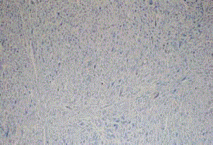
Figure 4A
CD117 negativity in malignant cells × 100.
Figure 4B
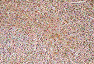
Figure 4B
Strong vimentin positivity in malignant cells × 100.
Discussion and Conclusion
Cancer at the colonic location is mainly of epithelial origin whereas mesenchymal tumors are very rare and form a heterogeneous group. These tumors may present with abdominal pain, rectal or intraabdominal bleeding, weight loss, constipation, diarrhea, obstruction, tenesmus, and anorexia, similar to epithelial tumors [2-4]. Their clinical findings and macroscopic appearances also resemble epithelial tumors. However, they can be distinguished by pathology examination and immunohistochemical staining. SMA, desmin, vimentin, S-100, neuron-specific enolase, CD34 and CD117 are used for the immunohistochemical differential diagnosis of primary mesenchymal tumors. Pleomorphic sarcomas are immunohistochemically negative for S100, desmin, SMA, and CD117 [5,6]. As in our case, focal CD68 positivity is present with generally strong vimentin positivity and low specificity [7,8]. They may suggest gastrointestinal stromal tumors as a site and appearance. However, unlike pleomorphic sarcomas, gastrointestinal stromal tumors are usually positive for S100 and CD117 [9]. The optimal diagnostic approach and treatment of primary mesenchymal tumors is not clear due to the small number of published cases. If a tumor is resectable, complete surgical resection is recommended, as in colonic epithelial tumors [5]. In unresectable or stage 4 patients, tumor regression may be achieved by chemotherapy, radiotherapy, or chemoradiotherapy [10]. The most active chemotherapy regimen is AIM (doxorubicin/ifosfamide/mesna) [11]. Since our case was a metastatic case, palliative chemotherapy was administered following palliative surgery. The disease prognosis depends on its stage, and our case died six months after the diagnosis.
In conclusion, colonic pleomorphic sarcomas are very rare tumors. Although their clinical features resemble those of epithelial tumors, differential diagnosis of patients presenting with a colonic mass should include a variety of tumors.
References
- Bond JH. Update on colorectal polyps: management and follow-up surveillance. Endoscopy. 2003;35:35-40.
- Conlon KC, Casper ES, Brennan MF. Primary gastrointestinal sarcomas: analysis of prognostic variables. Ann Surg Oncol. 1995;2:26-31.
- Lee CH, Jan YJ, Chen JT, Ho WL, Tseng CH, Wang J. Colorectal mesencymal tumor: A clinicopathologic study of 25 cases. J Chin Med Assoc. 2005;68:291-8.
- Nishimura J, Morii E, Takahashi T, Souma Y, Nakajima K, Doki Y, et al. Abdominal soft tissue sarcoma: a multicenter retrospective study. In J Clin Oncol. 2010;15:399-405.
- Yaren A, Değirmencioğlu S, Çallı Demirkan N, Gökçen Demiray A, Taşköylü B, Doğu GG. Primary mesenchymal tumors of the colon: A report of three cases. Turk J Gastroenterol. 2014;25:314-8.
- Udaka T, Suzuki Y, Kimura H, Miyashita K, SuwakiT,Yoshino T. Primary malignant fibrous histiocytoma of the ascending colon: report of a case. Surg Today. 1999;29:160-4.
- Goldblum JR. An approach to pleomorphic sarcomas: can we sub classify, and does it matter? Mod Pathol. 2014;27Supp1:S39-46.
- Fletcher CDM, Bridge JA, Hogendoorn PCW, Mertens F, editors. IARC. Undifferentiated/unclassified sarcomas, in WHO Classification of Tumours of SoftTissue and Bones. France: Lyon; 2013.
- vanRoggen JFG, vanVelthuysen MLF, Hogendoorn PCW. The histopathological differential diagnosis of gastrointestinal stromal tumors. J Clin Pathol. 2001;54:96-102.
- Yoon SS, Chen Y-L, Kambadakone A, Schmidt B, DeLaney TF. Surgical placement of biologic mesh spacers prior to external beam radiation for retroperitoneal and pelvic tumors. Pract Radiat Oncol. 2013;3:199-208.
- Judson I, Verweij J, Gelderblom H, Hartmann JT, Schöffski P, Blay JY, et al. Doxorubicin alone versus intensified doxorubicin plus ifosfamide for first-line treatment of advanced or metastatic soft-tissue sarcoma: a randomised controlled phase 3 trial. Lancet Oncol. 2014;15:415-23.