Case Report
Morgagni Diaphragmatic Hernia in Children: One Center’s Experience
Saud Al Shanafey*
Department of Surgery, King Faisal Specialist Hospital and Research Centre, Saudi Arabia
*Corresponding author: Saud Al Shanafey, Department of surgery, King Faisal Specialist Hospital and Research Centre, Riyadh, Saudi Arabia
Published: 02 Jul, 2018
Cite this article as: Al Shanafey S. Morgagni Diaphragmatic Hernia in Children: One Center’s Experience. World J Surg Surgical Res. 2018; 1: 1021.
Abstract
Background: Morgagni hernia is a rare type of congenital diaphragmatic hernia in children. We reviewed our experience with this anomaly at a tertiary care institution.
Methods: A retrospective chart review was conducted from June 2001 to June 2017. All patients managed for Morgagni hernia were included. Demographic, clinical, and outcome data were collected and descriptive data were generated.
Results: 21 patients with Morgagni hernia were managed over that period. There were 16 males and 5 females with mean age of 25 months. 15 (76%) patients have Down syndrome; 9 of them (43%) had previous sternotomy. 5 right sided, 11 left sided and 5 bilateral. All repaired laparoscopically except one, and mean weight at surgery was 9.7 Kg. Mean operative time was 93 minutes, and there were no postoperative complications. Patients were fed on the 1st postoperative day and mean hospital stay was 3 days. Median follow up was 60 months. There was one recurrence after 1 year of repair which was managed laparoscopically.
Conclusions: Morgagni Hernia is feasible to laparoscopic repair with favorable outcome. Our data may suggest an association between Morgagni hernia and Down syndrome but it was not clear if it was congenital or secondary to poor healing post sternotomy.
Keywords: Morgagni hernia; Diaphragmatic hernia; Laparoscopy
Introduction
Congenital diaphragmatic hernia is a rare anomaly occurs in 1:5,000 live births, and 3% to 5% of them are of the Morgagni type [1]. Morgagni Diaphragmatic Hernia (MDH) is an anterior defect in the subcostosternal portion of the diaphragm. These defects are usually congenital, but acquired cases have been reported [2]. Compared to the most common congenital diaphragmatic hernia (the posterolateral bochdalek type) MDH has not received much attention in the literature. The latter might be due to its rarity, its benign indolent course, and its less deleterious effect on the patients. We have reviewed our experience at King Faisal Specialist Hospital and Research Center (Tertiary Care Institution) with the management of Morgagni Hernia over the last 8 years.
Methods
A retrospective chart review was conducted for patients managed for MH at our institution in the period from June 2001 to June 2017. Demographic, clinical, and follow up data were collected and descriptive data were generated.
There was no missing data. Two techniques were used for repair of MDH, the intracorporeal and the extracorporeal. The intracorporeal technique entails suturing the posterior edge of the defect to the anterior abdominal fascia laparoscopically from inside the peritoneal cavity. The extracorporeal technique entails U-shaped stitches placed transabdominally under laparoscopic guidance and incorporating the posterior edge of the defect and then exiting the peritoneal cavity transabdominally to be tied extracorporeally incorporating the anterior abdominal fascia. The stitches are typically placed via a small skin incision in the epigastrium. The latter technique has been reported previously [3]. The study was approved by the clinical research committee and the research ethics committee at our institution.
Results
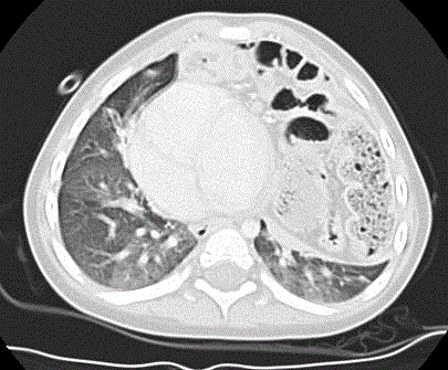
Twenty-one patients with Morgagni Hernia were managed over that period. There were 16 males and 5 females with a mean age at the time of repair of 25 months (range 8 to 68 months). Fifteen (76%) patients have Down syndrome; 9 of them (43%) had previous sternotomy. Nine patients presented with recurrent chest infections, while 12 were detected incidentally. All patients were diagnosed with Chest x-rays (Figure 1A and 1B), and 6 of them had CT scan for confirmation by referring services (Figure 2). Three were 5 right sided, 11 were left sided and 5 were bilateral. Mean weight at surgery was 9.7 Kg (range 4.6 Kg to 15.5 Kg), and mean operative time was 93 minutes (range 45-175 minutes). All repaired laparoscopically except one patient early in the series. Two patients had other procedures performed simultaneously with the repair of Morgagni Hernia. One had duodenal atresia (duodenoduodenostomy) and the other had gastro esophageal reflux disease (nissen fundoplication and gastrostomy tube insertion). The laparoscopic approach was conducted utilizing 3 trocars including the telescope port. Sizes of the trocars were 3.5 mm except in 4 patients where 5 mm trocars were utilized. Among the laparoscopic group, the 1st two patients were repaired utilizing the intracorporeal suturing technique, while the extracorporeal technique was utilized in the rest. Nineteen patients (including the open procedure) had their defects repaired primarily using interrupted 2.0 Silk sutures and 2.0 Proline in 1 patient. One patient had her defect repaired laparoscopically using Dacron patch to avoid tension. The latter had previous repair of a congenital Hiatal hernia. There were no postoperative complications. Patients were fed on the 1st postoperative day and mean hospital stay was 3 days (range 2-7 days). Median follow up was 60 months (range 10-100 months). There was one recurrence after 1 year of repair which was managed laparoscopically. The latter patient had a large hernia sac that extended to the apex of the right hemi thorax which was not respected primarily. During the repair of the recurrence, the hernia sac was partially respected and was managed utilizing the extracorporeal technique.
Figure 1A
Figure 1A
Chest X-ray (lateral view) shows a Morgagni Diaphragmatic Hernia with bowel content (arrows).
Figure 1B
Figure 2
Discussion
Morgagni diaphragmatic hernia is rare and may present early or late in life [2,4]. It is mostly asymptomatic and the defect is usually detected incidentally on a radiological exam performed for other purposes [5]. Chest x-ray is the most common diagnostic test, especially the lateral view. Patients usually present with respiratory or gastrointestinal symptoms [5,6]. Symptomatic patients mostly present with recurrent chest infections, and it is not unusual to detect the abnormality on a chest x-ray performed after the resolution of a chest infection [2,7].
Gastrointestinal presentation is less common and usually obstructive in nature due to incarceration of bowel loops (usually the colon) in the hernia sac [8,9]. In many cases the diagnosis is delayed due to its rarity and the non-specific presentation [2]. It is advocated that a high level of suspicion have to be exercised when assessing patients with unusual respiratory and gastrointestinal symptoms [2,4].
Surgical Repair of MDH is recommended on a semi-urgent basis, even for asymptomatic patients, to avoid associated morbidities [2,4]. Many approaches were described for MDH repair. Historically, open thoracic or abdominal (midline or transverse) techniques were used, but lately laparoscopic repair was described with high success rate [3,10,11]. Laparoscopic repair method also varies in the literature. Some described total intracorporeal primary repair [10], while others advocated a transabdominal sutures with extracorporeal knots with comparable results. There are no controlled studies to compare the intracorporeal and the extracorporeal approaches [3]. Prosthetic patch was used primarily for repair by many authors to avoid tension [4,10-14], and some have used the prosthetic patch to augment the primary repair [15]. We and others favor excision of the hernia sac to avoid recurrence [16] while others do not feel it is necessary [17,18]. In our series, the single recurrence occurred in the patient who had a very large hernia sac extending to the apex of the right hemithorax which made its resection difficult. This patient had his sac partially removed in the redo procedure and has no recurrence 48 months after. We believe that a laparoscopic approach should be utilized in these cases given its safety, feasibility, and the many potential advantages that the minimally invasive technique offers to patients [16,18]. These include faster recovery, less pain, shorter hospitalization, and superior cosmetic results. We also advocate a primary repair with non-absorbable sutures to avoid complications of the prosthetic patch and due to the fact that primary repair was effective in our experience and others [3]. If the defect is large and significant tension is anticipated from primary repair, utilization of prosthetic patch to avoid recurrence is recommended. One of our patients, who had a previous repair of a congenital Hiatal hernia, required Dacron patch to avoid tension.
Most of the reports concerning MDH are case reports and case series with emphasis on the rarity of the anomaly and the surgical technique used for its correction. In our series, 75% of our patients were known with Down syndrome. Of patients with Down syndrome, 56% with congenital heart disease and presented after sternotomy procedures for correction of the heart anomaly. This may reflect the nature of our institution as a tertiary care center with major cardiac surgery service. Moreover, patients post sternotomy performed for heart procedures end up with chest drains inserted in the subcostosternal area where MDH occurs. We could speculate that poor tissue healing in that area may have contributed to the development of MDH in these patients. The latter is supported by the fact that tissue healing in Down syndrome patients is impaired due to altered cellular immunity [19]. On the other hand, many reports have suggested an association between MDH and Down syndrome [7,20-22] and 6 of our patients have Down syndrome without previous sternotomy. In addition, despite the high number of patients with normal chromosomes managed for congenital heart disease surgically with sternotomy at our institution (around 500 procedures annually), we have not encountered a single case of MDH over the same period. Our findings may suggest an association between MDH and Down syndrome rather than defected healing post-sternotomy.
References
- Snyder WH, Greany EM. Congenital diaphragmatic hernias: 77 consecutive cases. Surgery. 1965;57:567-88.
- Alsalem AH. Congenital hernia of Morgagni in infants and children. J Pediatr Surg. 2007;42(9):1539-43.
- Azzie G, Maoate K, Beasley S, Retief W, Bensoussan A. A simple technique of laparoscopic full-thickness anterior abdominal wall repair of retrosternal (Morgagni) hernias. J Pediatr Surg. 2003;38(5):768-70.
- Vyhnanek M, Rygl M, Snajdauf J, Skaba R, Kyncl M. Morgagni diaphragmatic hernia in childhood. Rozhl Chir. 2006;85(10):494-7.
- Pokorny WJ, McGill CW, Harberg FJ. Morgagni hernias during infancy: Presentation and associated anomalies. J Pediatr Surg. 1984;19(4):394-97.
- Berman L, Stringer D, Ein SH, Shandling B. The late presenting pediatric Morgagni hernia: A benign condition. J Pediatr Surg. 1989;24(10):970-2.
- Parmar RC, Tullu MS, Bavdekar SB, Borwankar SS. Morgagni hernia with Down syndrome: a rare association—case report and review of literature. J Postgrad Med. 2001;47(3):188-90.
- Barut I, Tarhan OR, Cerci C, Akdeniz Y, Bulbul M. Intestinal obstruction caused by a strangulated Morgagni hernia in an adult patient. J Thorac Imaging. 2005;20(3):220-2.
- Becini L, Pampaloni F, Taddei G. Intestinal occlusion caused by strangulated morgagni-larry hernia: clinical case and review of the literature. Chir Ital. 2001;53(3):415-9.
- Durak E, Gur S, Cokmez A, Atahan K, Zahtz E, Tarcan E. Laparoscopic repair of Morgagni hernia. Hernia. 2007;11(3):265-70.
- Dutta S, Albanese CT. Use of prosthetic patch for laparoscopic repair of Morgagni diaphragmatic hernia in children. J Laparoendosc Adv Surg Tech A. 2007;17(3):391-4.
- Filipi CJ, Marsh RE, Dickason TJ, Gardner GC. Laparoscopic repair of a Morgagni hernia. Surg Endosc. 2000;14(10):966-7.
- Tan YW, Banerjee D, Cross KM, De Coppi P, Blackburn SC, Rees CM, et al. Morgagni hernia repair in children over two decades: Institutional experience, systematic review, and meta-analysis of 296 patients. J Pediatr Surg. 2018:S0022-3468(18)30248-3.
- Yavuz N, Yigitbasi R, Sunamak O, As A, Oral C, Erguney S. Laparoscopic repair of Morgagni hernia. Surg Laparosc Endosc Percutan. Tech 2006;16(3):173-6.
- Dapri G, Himpens J, Hainaux B, Roman A, Stevens E, Capelluto E, et al. Surgical technique and complications during laparoscopic repair of diaphragmatic hernias. Hernia. 2007;11(2):179-83.
- Sahnoun L, Ksia A, Jouini R, Maazoun K, Mekki M, Krichene I, et al. Morgagni hernia in infancy: report of 7 cases. Arch Pediatr. 2006;13(10):1316-9.
- Akbiyik F, Tiryaki TH, Senel E, Mambet E, Livanelioglu Z, Atayurt H. Is hernial sac removal necessary? Retrospective evaluation of eight patients with Morgagni hernia in 5 years. Pediatr Surg Int. 2006; 22(10):825-7.
- Horton JD, Hofmann LJ, Hetz SP. Presentation and management of Morgagni hernias in adults: a review of 298 cases. Surg Endosc. 2008;22(6):1413-20.
- Spina CA, Smith D, Korn E, Fahey JL, Grossman HJ. Altered cellular immune functions in patients with Down syndrome. Am J Dis Child. 1981;135(3):251-5.
- Cigdem MK, Onen A, Okur H, Otcu S. Associated malformations in Morgagni hernia. Pediatr Surg Int. 2007;23(11):1101-3.
- Harris GJ, Soper RT, Kimura KK. Foramen of Morgagni hernia in identical twins: is this an inheritable defect? J Pediatr Surg. 1993;28(2):177-8.
- Honore LH, Torfs CP, Curry CJ. Possible association between the hernia of Morgagni and trisomy 21. Am J Med Genet. 1993;47(2):255-6.


