Research Article
Comparison between Indocyanine-Green and Fluorescein for Cerebrovascular Surgery
Atsushi Saito*
Department of Neurosurgery, Sendai Medical Center, Japan
*Corresponding author: Atsushi Saito, Department of Neurosurgery, Sendai Medical Center, 2-8-8 Miyagino, Miyagino-ku, Sendai, Miyagi, 0300910, Japan
Published: 28 Jun, 2018
Cite this article as: Saito A. Comparison between Indocyanine-Green and Fluorescein for Cerebrovascular Surgery. World J Surg Surgical Res. 2018; 1: 1017.
Abstract
Purpose: Intraoperative Fluorescent Angiography (IFA) with two types of fluorescent dyes of Fluorescein (FS) and Indocyanine Green (ICG) is a useful tool for cerebrovascular surgery. We compared the two fluorescent dyes on the points of detection of perforators and application bypass surgery.
Methods: Intraoperative fluorescent angiography was performed with intraoperative observatory modules of microscope of OPMI PENTERO900 including two types of induction light for both of FS and ICG to evaluate the same fluorescent operative field for them. Fluorescent dyes of 250 mg of FS and ten times dilution of ICG were intravenously administered and quantitative analysis was performed with ROIs of software for movie analyzer. Objects for evaluation of perforators were 17 cases of cerebral aneurysms treated in our department from December 2015 to May 2016.
Results: Detection of perforators was evaluated in 6 cases of ruptured aneurysms and 11 cases of unruptured aneurysms. Location of aneurysms was as follows: 6 cases in anterior communicating artery, 4 cases in internal carotid artery-posterior communicating artery, and 7 cases in middle cerebral artery. Fluorescent intensity of perforators were evaluated with hypothalamic artery, lenticulostriate artery and branches from posterior communicating artery and that of main trunks was evaluated as objects with A1, A2, M1, and M2 portions. Averaged fluorescent intensity of FS was 78.3 ± 25.8 in perforators and 117.4 ± 22.2 in main trunks. Averaged fluorescent intensity of ICG was 29.2 ± 14.0 in perforators and 76.2 ± 40.0 in main trunks. The ratio of averaged fluorescent intensity to maximum fluorescent intensity, which indicated contrast of vascular structure on fluorescent angiography, was 2.97 times in FS and 1.98 times in ICG. Fluorescent angiography was applied to a case associated with postoperative hyper perfusion after superficial temporal artery-middle cerebral artery bypass. Fluorescent intensity of cortex was markedly higher in FS than that of ICG just after bypass.
Conclusions: Fluorescein was superior to detect perforators during clipping compared with ICG on both points of fluorescent intensity and contrast. Fluorescent angiography with FS might be superior to predict hyper perfusion after bypass surgery.
Introduction
A variety of intraoperative monitoring methods were applied to cerebrovascular surgery, especially to evaluate ischemic insult during clipping of aneurysms, such as temporary occlusion of parent artery, injury of perforators. Electrophysiological diagnosis by motor evoked potential and ultrasonic blood flow monitors and intraoperative digital subtraction angiography are useful methods, but all have limitation. Intraoperative fluorescent angiography with Fluorescein (FS) and Indocyanine Green (ICG) is one of simple monitoring methods and recently applied to broader field of microsurgery. However, comparative studies of the two fluorescent dyes are few. We compared FS and ICG on the points of detection of perforators during clipping and application to bypass surgery using microscope including induction lights for both fluorescent dyes.
Methods
Objects were 17 cases of aneurysmal clipping treated in our department from 2015 to 2016. OPMI Pentero was used and two induction lights for FS and ICG were included intraoperative observation module. The same operative field could be prepared for fluorescent angiography using the different fluorescent dyes. Fluorescein and ICG were administered intravenously before and after clipping. Fluorescein and ICG were administered intravenously to evaluate patency after bypass surgery. Five milliliter of ICG diluted 10 times was administered after clipping or bypass. Thereafter, 5 ml of FS diluted 10 times was administered. Intra arterial flow of fluorescent dyes was observed under microscope and saved as movie. Movie files were saved as AVI files and regions of interest were set on cerebral arteries, veins and cortical surfaces and changes of fluorescent intensity were quantitatively analyzed with movie analyzer software, ROIs. On detection of perforators after aneurysmal clipping, regions of interest were set on both main trunks and perforators. Main trunks were A1 and A2 portions of anterior cerebral artery, or M1 and M2 portions of middle cerebral artery. Targets of perforators were hypothalamic artery, lenticulostriate artery; perforators branched from posterior communicating artery. A number of perforators could be detected under microscope and on saved movie were counted as detectable perforators and the data was compared between FS and ICG.
To evaluate intensity of contrast in fluorescent angiography, ratio of averaged fluorescent intensity of the target vessel to the maximum fluorescent intensity of the neighbor main trunk was used. Averaged fluorescent intensity was defined as averaged intensity value from the initial point of detection of fluorescent light to the last point of detection of fluorescent light via reaching peak and fading. To calculate the ratio of averaged fluorescent intensity, averaged intensity was divided by the maximum fluorescent intensity of the neighbor main trunk.
Table 1
Table 1
Location and characteristics of aneurysms and target arteries.
Acom: Anterior Communicating Artery; MCA: Middle Cerebral Artery; ICA: Internal Carotid Artery; AN: Aneurysm
Table 2
Table 3
Table 3
Fluorescence intensity of main trunks on fluorescence cerebral angiography.
ICG: Indocyanine Green
Table 4
Table 4
Fluorescence intensity of perforators on fluorescence cerebral angiography.
ICG: Indocyanine Green
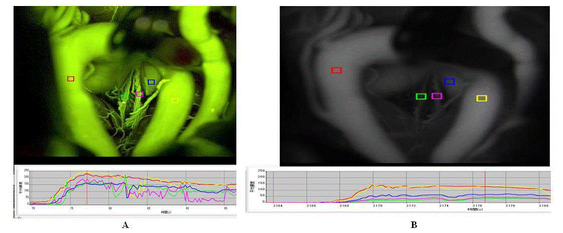
Figure 1A and 1B
Figure 1A and 1B
Representative image of clipped anterior communicating artery aneurysm on fluorescence cerebral angiography after intravenous administration
of fluorescein (Figure 1A) and indocyanine green (Figure 1B). Various colored squares indicated regions of interest for quantitative analysis of fluorescence intensity
(upper images of Figure 1A and 1B. Time-dependent changes of fluorescence intensity showed curve of each region of interest (lower graphs of Figure 1A and 1B).
Results
Detection of perforators during clipping of cerebral aneurysms Perforators was detected with intraoperative fluorescent angiography in 6 cases of ruptured aneurysms and in 11 cases of unruptured aneurysms (Table 1). Locations of the aneurysms were 6 cases in anterior communicating artery, 7 cases in middle cerebral artery and 4 cases of internal carotid artery. Objects for detection of perforators with postoperative video were performed 43 main trunks and 34 perforators detected under microscope during clipping. In fluorescent angiography, all 34 perforators were detected in both under microscope and postoperative video (Table 2). In ICG angiography, 30 perforators were detected comparing with those detected under microscope (Table 2). Detection of perforators in fluorescent angiography with fluorescein was superior to in that with ICG. Difficult cases in ICG angiography were all perforators from posterior communicating artery.
In fluorescent contrast data, averaged intensity was 117.4% and the maximum intensity was 240.9 % in fluorescent angiography with fluorescein in main trunks (Table 3). On the other hand, averaged intensity was 76.2% and the maximum intensity was 126.7% in fluorescent angiography with ICG. In perforators, averaged intensity was 78.3% and the maximum intensity was 213.9% in fluorescent angiography with fluorescein (Table 4). Averaged intensity was 29.2% and the maximum intensity was 60.3% in fluorescent angiography with ICG. Contrast data in fluorescent angiography with fluorescein was superior to that in that with ICG.
Discussion
Evaluation of ischemic insults in main trunks and perforators during cerebrovascular surgery is important for safer surgery and a variety of intraoperative monitoring methods have been developed [1,2]. Suzuki et al. [1] reported benefits and defects of some monitoring as follows: sonographic evaluation is available for main trunks, but is not suitable for small vessels such as perforators, and tissue blood flow meter shows quantitative value on cortical surface, but data is unstable on each measurement. Electrophysiological monitoring has a benefit for neurological function, but it cannot evaluate targeted tissue except for neurological circuit. Intraoperative x-ray based angiography has risks of complications surroundings of catheter procedures and higher invasiveness [1].
Fluorescent angiography has been reported as a beneficial method, but each fluorescent dye has effect on detection of vessels and knowledge of characters contributes to better intraoperative evaluation and safer cerebrovascular surgery.
The maximum wave length of fluorescein and ICG is 465-490/520-530 nm and 805/835 nm, respectively and serial trials of fluorescent angiography by intravenous administration of these dyes do not interfere with each detection [2,3,4]. Intravenous administration of fluorescent dyes has some disadvantages as follows: First, it needs many amounts of fluorescent dyes at each injection and wash-out of fluorescent intensity delays. Fluorescent dyes remain in vessels even 5-6 minutes after injection. Intravenous fluorescent angiography is not suitable for evaluation of venous circulatory disturbance. Second, Intravenous fluorescent dyes are diluted via cardio-pulmonary circulation to intracranial arteries. Concentration of dyes decreases at the time of reaching cerebral arteries and fluorescent intensity elevates slowly [1]. Kuroda et al. [5] tried intra arterial injection of fluorescent dyes during aneurysmal surgery and emphasized usefulness on clipping for the following reasons: Quality of fluorescent images was superior to intravenous injection and rapid wash-out of dyes enabled to repeat serial angiography [5]. Intravenous injection has advantage of simple procedure comparing with intra arterial injection and should be applied to intraoperative evaluation based on these points.
Intraoperative detection of perforators is important and has influences on postoperative prognosis. Fluorescent angiography has advantages to evaluate not only anatomical and morphological vascular structures, but also visualization of blood flow itself [2,6-8]. We examined both fluorescent intensity and contrast in order to evaluate visualization of two kinds of fluorescent dyes. To our knowledge, there was no report of decisive methods to evaluate the contrast of fluorescent dyes. Ratio of averaged fluorescent intensity of targeted vessels to the maximum fluorescent intensity of the neighbor main trunks was defined as a marker to evaluate the visualization in this study. Fluorescent images of perforators by fluorescein were superior on fluorescent intensity and contrast to those of ICG. Detection of perforators might be affected by depth of targets and existence of hematoma surrounding with vessels. Characteristics and wash-out of each fluorescent dye should be considered in repeat of fluorescent angiography and association with other monitoring methods such as Doppler sonography and motor evoked potential might be also useful.
In our study, following limitation should be considered. The number of objects was small and retrospective study in a single institution. Fluorescent intensity was evaluated with recorded video images. However, there might be slight difference between images under microscope and recorded video images in intensity, clearness and contrast. It was difficult to evaluate superiority on detection of perforators under complete same condition. Especially, quality of images in ICG might be affected in deeper region. Intensity of background also might have influences in fluorescent intensity on vessels. Precise examination considering depth of target vessels and recorded images will be required in future studies.
References
- Suzuki K, Ichikawa T, Wayanabe Y. Evaluation of cortical blood flow by intra-arterial fluorescence cerebral angiography: Comparison between indocyanine-green and fluorescein in three cases. Surg Cereb Stroke. 2014;42(3):207-13.
- Kyouichi S, Namio K, Tatsuya S, Masato M, Tsuyoshi I, Ryoji M. Confirmation of blood flow in perforating arteries using fluorescein cerebral angiography during aneurysm surgery. J Neurosurg. 2007;107(1):68-73.
- Rey-Dios R, Cohen-Gadol AA. Technical principles and neurosurgical applications of fluorescein fluorescence using a microscope-integrated fluorescence module. Acta Neurochir. 2013;155(4):701-6.
- Woitzik J, Peña-Tapia PG, Schneider UC, Vajkoczy P, Thomé C. Cortical perfusion measurement by indocyanine-green videoangiography in patients undergoing hemicraniectomy for malignant stroke. Stroke. 2006;37(6):1549-51.
- Kuroda K, Kinouchi H, Kanemaru K, Nishiyama Y, Ogiwara M, Yoshioka H, et al. Intra-arterial injection fluorescein videoangiography in aneurysm surgery. Neurosurgery. 2013;72(2):141-50.
- Raabe A, Beck J, Gerlach R, Zimmermann M, Seifert V. Near-infrared indocyanine green video angiography: A new method for intraoperative assessment of vascular flow. Neurosurgery. 2003;52(1):132-9.
- Raabe A, Nakaji P, Beck J, Kim LJ, Hsu FP, Kamerman JD. Prospective evaluation of surgical microscope-integrated intraoperative near-infrared indocyanine green videoangiography during aneurysm surgery. J Neurosurg. 2005;103(6):982-9.
- Wrobel CJ, Meltzer H, Lamond R, Alksne JF. Intraoperative assessment of aneurysm clip placement by intravenous fluorescein angiography. Neurosurgery. 1994;3(5):970-3.


