Case Report
Topical Application of Adipose-Derived Mesenchymal Stem Cells in the Treatment of a Non-Healing Ulcer in a Leper: A Case Report - A New Option for Treatment of Extremely Chronic Ulcer
Lam PK1,2, Winne Lo3, Cindy SW Tong1,2, Lo KY1,2, Ping Chook3, Paul BS Lai1,2, Poon WS1,2 and Leung PC3*
1Department of Surgery, Chinese University of Hong Kong, Hong Kong
2Chow Tai Fook-Cheng Yu Tung Surgical Stem Cell Research Center, Chinese University of Hong Kong, Hong Kong
3Institute of Chinese Medicine, Chinese University of Hong Kong, Hong Kong
*Corresponding author: Leung PC, Institute of Chinese Medicine, Prince of Wales Hospital, Chinese University of Hong Kong, Hong Kong
Published: 27 Jun, 2018
Cite this article as: Lam PK, Winne Lo, Cindy SW Tong, Lo KY, Ping Chook, Paul BS Lai, et al. Topical Application of Adipose-Derived Mesenchymal Stem Cells in the Treatment of a Non-Healing Ulcer in a Leper: A Case Report - A New Option for Treatment of Extremely Chronic Ulcer. World J Surg Surgical Res. 2018; 1: 1016.
Abstract
Neuropathic ulceration in the foot is one of the most serious complications in leprosy. The healing is much impaired because of chronic inflammation, deformation and neurological impairment. Current wound management protocols commonly fail to heal. The multipotency paracrine and immunomodulatory functions of Mesenchymal Stem Cells (MSC) enable them to offer a novel regenerative therapy to expedite the healing of lepromatous ulcer. In this case study, the 20 years chronic ulcer on the deformed foot of a leper healed after the left sole ulcer of a patient with a history of leprosy healed after topical application of expanded MSC. The ulcer had no recurrence up to 15 months after healing. No adverse defects were detected. The result of this pilot case is highly impressive. Experimental works to explore the mechanisms of healing are underway.
Keywords: Stem cell; Non-healing ulcer; Topical application
Introduction
Leprosy is an infectious disease caused by mycobacterium leprae affecting peripheral nerves resulting in deformities and loss of sensation. Moreover leprosy has a tropism for macrophages and Schwann cells [1]. Neuropathic ulceron the sole is one of the most common complications after the infection is controlled. Approximately 10% to 20% in leprosy patients develop chronic ulcers [2]. The impairment of the wound healing is primarily due to the chronic inflammation, loss of sensation, deformities and markedly decreased healing power. The multipotency, safety of Mesenchymal Stem Cells (MSCs) in clinical trials and the impressive paracrine and immunomodulatory properties of MSC have prompted us to try healing extremely chronic ulcers like lepromatous ulcer with MSCs.
Case Presentation
The patient was a 71-years old man with a history of leprosy since 40 years ago. Infection has been controlled and he is maintained on anti-leprosy medication. He developed a chronic ulcer on the left sole since 20 years ago. On presentation, the ulcer was 4 cm × 2.5 cm with heavy growth of Pseudomonas aeruginosa. The foot was markedly deformed and shrunken to about 50% of a normal foot (Figure 1).
With the approval from the Clinical Research Ethics Committee and informed consent from the patient, about 1 gm of subcutaneous adipose tissue was obtained around the umbilical site under local anesthesia. The adipose tissue was washed extensively with phosphate buffered saline and then digested with 0.1% collagenase (type I; Sigma-Aldrich) for 30 minutes at 37°C with gentle agitation. After filtration through 100-μm mesh filter, the filtrate was washed three times and completely suspended in Dulbecco’s Modified Eagle’s Medium (DMEM) supplemented with 10% fetal bovine serum. The cultures were maintained in a humidified atmosphere of 5% CO2. MSCs at cell passage 3 to 5 were used for cell transplant. The MSCs demonstrated the potential to differentiate into adipocytes, chondroblasts and osteoblasts under specific in vitro conditions. Flow cytometry showed ADMSCs expressed CD13, CD29, CD44, CD90, CD105 and CD166 but not CD34, CD31 and CD45 (Figure 2).
All cytotoxic topical antimicrobial solutions were discontinued two days before the cell transplant. About 2 MSCs × 107 MSCs suspended in 500 ul PBS was topically applied to the wound surface. A thin layer of fibrin (Tisseel, Baxter) was sprayed to fix the MSCs in position. The wound was then dressed with a non-adherence dressing (Metipel) followed by Vaseline gauze (Adaptic). The wound was examined once a week. MSCs were grafted every 3 to 4 weeks until it healed. There was persistent Pseudomonas aeruginosa. The ulcer completely healed after 14 times of topical application of MSCs in 6 months (Figure 3). There was no complication arising from the topical MSCs and no recurrence of the ulcer 15 months after the healing.
Figure 1
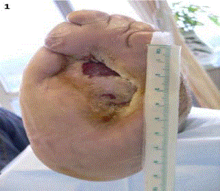
Figure 1
A non-healing lepromatous ulcer.
Figure 2
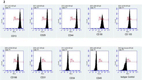
Figure 2
Phenotype of the adipose tissue-derived MSCs.
Figure 3
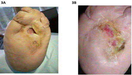
Figure 3
The non-healing lepromatous ulcer- (A) 3 months after 10 MSC transplants and (B) healed after 14 MSC transplant within 8 months.
Discussion
Wound healing has four distinct phases: Homeostasis, inflammation, granulation tissue formation and matrix remodeling involving a number of well-orchestrated interactions among the infiltrating and resident cells, mediators, growth factors and cytokines. Formation of new blood vessels within the ulcer bed (i.e. granulation tissue) is one of the crucial steps of wound healing. Under normal circumstances, angiogenesis and vasculogenesis of the granulation tissue are regulated by proangiogenic growth factors such as Vascular Endothelial Growth Factor (VEGF) [3]. Once the blood supply is sustained within the wound bed, re-epithelialization and matrix remodeling can proceed to develop a stable framework of collagen and elastin fibers.
Leprosy is a chronic infectious disease caused by Mycobacterium leprae which is an obligate intracellular neurotrophic bacteria infecting the Schwann cells. The damage of peripheral nerves in the limbs results in sensory loss and motor impairments. The pathogenesis of lepromatous ulcer is enhanced by the continuous breakdown of the plantar tissue by the abnormal high pressure exerted on the sole of the deformed foot during persistent walking. Vulnerability to infection further delays the healing of lepromatous ulcer.
MSCs are multipotent adult stem cells present in different kinds of tissues such as bone marrow, adipose tissue, and capable of self-renewal and differentiation into mature cells of various lineages [4]. The ease of isolation and efficient expansion in vitro make adipose tissue-derived MSCs an excellent candidate for cell therapy. Growing evidence show that differentiation of transplanted MSCs into phenotype of epithelium is not considered as the major recovery mechanism to accelerate cutaneous wound [5,6]. The release of a variety of cytokines from MSCs is thought to play a significant role in tissue repair though suppression of inflammation and apoptosis, as well as stimulating angiogenesis and re-epithelialization. In fact, the beneficial effects of MSCs on wound healing have been demonstrated in a variety of preclinical and clinical studies [7-9]. MSCs enhance re-epithelialization, angiogenesis and granulation tissue formation. MSCs have been administrated by intramuscular/subcutaneous injection or topical application onto the recipient wounds of ulcers. In this case study, topical application onto the ulcer bed was performed. In order to sustain the continuous effects of MSCs, a film of fibrin was sprayed on top to prevent the escape of MSCs under the dressings.
To our best knowledge, this is the first report on the use of autologous MSCs to treat a lepromatous ulcer. Although the outcome of this preliminary clinical trial is promising, the role of MSCs in the healing of lepromatous ulcer is yet to be determined. In the meantime, we are recruiting more cases and would investigate whether the transplanted MSCs directly differentiate and engraft into the granulation tissue, or just mediate the healing process through their paracrine effects. Experimental work on animals is underway for the exploration on the mechanisms of healing using MSCs.
References
- Shimoji Y, Ng V, Matsumura K, Fischetti VA, Rambukkana A. A 21-kDa surface protein of Mycobacterium leprae binds peripheral nerve lamin-2 and mediates Schwann cell invasion. Proc Natl Acad Sci U S A. 1999;96(17):9857-62.
- Rodrigues LC, Lockwood DNJ. Leprosy now: Epidemiology, progress, challenges and research gaps. Lancet Infect Dis. 2011;11(6):464-70.
- Wu Y, Chen L, Scott PG, Tredget EE. Mesenchymal stem cells enhance wound healing through differentiation and angiogenesis. Stem Cells. 2007;25(10):2648-59.
- Schaffler A, Buchler C. Concise review: Adipose tissue-derived stromal cells-basic and clinical implications for novel cell-based therapies. Stem Cells. 2007;25(4):818-27.
- Chen JS, Wong VW, Gurtner GC. Therapeutic potential of bone marrow-derived mesenchymal stem cells for cutaneous wound healing. Front Immunol. 2012;3:192.
- Hocking AM, Gibran NS. Mesenchymal stem cells: Paracrine signaling and differentiation during cutaneous wound repair. Exp Cell Res. 2010;316(14):2213-9.
- Mulder GD, Lee DK, Jeppesen NS. Comprehensive review of the clinical application of autologous mesenchymal stem cells in the treatment of chronic wounds and diabetic bone healing. Int Wound J. 2012;9(6):595-600.
- Wu Y, Huang S, Enhe J, Fu X. Insights into bone marrow-derived mesenchymal stem cells safety for cutaneous repair and regeneration. Int Wound J. 2012;9(6):586-94.
- Papa ND, Luca GD, Sambataro D, Zaccara E, Maglione W, Gabriella A, et al. Regional implantation of autologous adipose tissue-derived cells induces a prompt healing of long-lasting indolent digital ulcers in patients with systemic sclerosis. Cell Transplant. 2015;24(11):2297-305.