Case Report
Umbilical Epidermoid Cyst Associated with an Urachal Remnant: A Case Report and Review of Literature
Volkan S Erikci*
Department of Pediatric Surgery, Saglık Bilimleri University, Tepecik Training Hospital, Turkey
*Corresponding author: Volkan S Erikci, Department of Pediatric Surgery, Saglık Bilimleri University, Tepecik Training Hospital, İzmir, Turkey
Published: 21 May, 2018
Cite this article as: Erikci VS. Umbilical Epidermoid Cyst Associated with an Urachal Remnant: A Case Report and Review of Literature. World J Surg Surgical Res. 2018; 1: 1010.
Abstract
Epidermoid Cysts (EC) are uncommon in children. They can occur on anybody surface but the most common sites of involvement include face, neck and trunk. Umbilicus is a rare site of involvement which sometimes leads to misdiagnosis. Herein we report a case of umbilical EC associated with an atretic urachal remnant in a 5-year-old boy. The patient is discussed under the light of relevant literature. Umbilical EC should be kept in mind in the differential diagnosis of a child with umbilical mass and awareness of this lesion can avoid its misdiagnosis and inappropriate treatment.
Keywords: Epidermoid cyst; Umbilicus; Urachal remnant
Introduction
An EC is a benign cyst usually found on the skin of face, neck and trunk [1]. It has been postulated that it derives from ectodermal tissue lined by the squamous epithelium devoid of skin appendages. They may occur anywhere on the body except palms of the hands and soles of the feet [2,3]. Umbilicus is a rare site of involvement in EC and when located in the umbilicus, EC usually leads to misdiagnosis including umbilical hernia, umbilical polyp and remnants of umbilicus and urachus [1,4-6]. In this report, a 5-year-old boy with an umbilical EC associated with an atretic urachal remnant is presented and discussed. It is also aimed in this report to review the presentation and management of these patients. Awareness of this lesion can avoid its misdiagnosis and inappropriate treatment.
Case Presentation
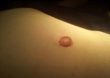
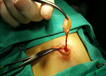
An otherwise healthy 5-year-old boy presented with a swollen was in his umbilicus (Figure 1). The history of the patient revealed that this mass had been present since birth. There was no pain, no discharge from umbilicus, but an increase in size of swelling was noted by his parents and this prompted the family to seek a medical assistance. Physical examination revealed a subcutaneous mass without tenderness, soft in consistency, non-reducible and mobile in all directions. Ultrasound of the mass and abdomen revealed a subcutaneous mass without a connection to the abdominal cavity or internal organs. Surgical intervention under general anesthesia revealed a lipomatous mass connected with an atretic urachal remnant and total excision was performed with a circumumbilical incision (Figure 2). Histopathological assessment revealed an EC associated with an atretic and obliterated urachal remnant. The child made a good post-operative recovery and was discharged home well the following day. The infant is otherwise well with no significant past medical history.
Figure 1
Figure 2
Discussion and Conclusion
Derived from ectodermal tissue, an EC is a benign lesion commonly found on the skin. It
usually involves the scalp, face, neck, back and trunk [7]. It is usually asymptomatic and when it
ruptures, and its keratin content faces with the dermis, foreign body reaction may be incited [8].
Usual reasons for seeking medical assistance in these patients include infection and an increase in
size producing damage to adjacent anatomical structures [9]. Increase in size of umbilical mass in
the presented case was the reason for his family to seek for medical aid. Differential diagnosis of EC
includes dermoid cysts, lipomas and pilomatrixomas [4].
Umbilicus is an uncommon site for EC and usually leads to a misdiagnosis. Literature on this
issue is scarce and to our knowledge only a 9-year-old boy with an umbilical EC above fascia has been
reported previously [5]. Our case is unique in that, to our knowledge, he is the youngest patient with
umbilical EC reported in the English language literature. It has also been reported that umbilical EC
may also occur following abdominoplasty [10,11]. The differential diagnosis of umbilical EC include umbilical hernia, umbilical cord hernia, umbilical polyp, urachal
remnant in children, and umbilical adenoma, Sister Mary Joseph
nodule, caput medusa and desmoid in adults [1,4].
The urachus is a connection between the fetal bladder and the
allantois. There is a wide spectrum in the presentation of Urachal
Remnants (UR) including patent urachus, urachal sinus, urachal
diverticulum, urachal cyst and atretic UR or urachal chorda and there
is no consensus on the management of these lesions [6]. Although it
is stated that asymptomatic and non-specific atretic URs can be safely
managed non-operatively and that these will often resolve without
intervention, it is generally admitted that URs should be resected as
chronic exposure to urinary stasis, infection and inflammation in
the remnant may predispose to urachal carcinoma [6]. Umbilical
appearance may differ in patients with UR. Although an incidence of
9.7% abnormal appearance of the umbilicus on examination has been
reported previously, to our knowledge, there is no case with umbilical EC associated with a UR. Besides being the youngest patient in the
English language literature having umbilical EC, the presented case in
this report is unique in that although preoperatively it was not known
that the patient had an associated UR, during surgical intervention,
the umbilical EC was found to be associated with an atretic urachal
remnant which was confirmed histopathologically after total excision
of the mass.
In conclusion, slow growing and often painless ECs rarely cause
problems or need treatment. When located in uncommon sites such
as umbilicus as in the presented case, EC may produce diagnostic
dilemma leading to inappropriate treatment and should be resected.
The presented case shows that umbilical EC and an atretic UR may
coexist in the same patient. This report adds to literature that these
two entities can be seen in the same patient and the physicians and
radiologists should be aware of this rather rare clinical entity and
appropriate surgical intervention is paramount.
References
- Padasali PS, Ravikumara G, Sundeep VK, Shankaregowda VS. A rare case of umbilical epidermoid cyst. Surgery Curr Res. 2015;5:1-2.
- Klin B, Ashkenazi H. Sebaceous cyst excision with minimal surgery. Am Fam Physician. 1990;41:1746-8.
- Barnes L, editor. Surgical pathology of the head and neck. 3rd ed. New York: Informa Healthcare, USA. 2008.
- Hattori NA, Tanaka T, Mukai K, Kish K. Congenital epidermal cyst of the umbilicus: a case report. Modern Plast Surg. 2013;3:128-9.
- McClenathan JH. Umbilical epidermoid cyst: an unusual cause of umbilical symptoms. Can J Surg. 2002; 45:303-4.
- Naiditch JA, Radhakrishnan J, Chin AC. Current diagnosis and management of urachal remnants. J Pediatr Surg. 2013;48:2148-52.
- Kang SG, Kim CH, Cho HK, Park MY, YJ Lee, MK Cho. Two cases of giant epidermal cyst occurring in the neck. Ann Dermatol. 2011;23:135-8.
- Matsumono K, Okabe H, Ishizawa M, Egawa M. Intermetatarsophalangeal bursitis induced by a plantar epidermal cyst. Clin Orthop Relat Res. 2001;385:151-6.
- Basterzi Y, Sari A, Ayhan S. Giant epidermoid cyst on the forefoot. Dermatol Surg. 2002;28:639-40.
- Andreadis AA, Samson CM, Szomstein S, Newman IM. Epidermal inclusion cyts of the umbilicus following abdominoplasty. Plast Surg Nurs. 2007;27:202-5.
- Camenisch CC, Heden P. Umbilical epithelial cyst in secondary abdominoplasty: case report. Aesthetic Plast Surg. 2012;36:83-7.


