Research Article
3T MRI in the Evaluation of Acute Appendicitis in the Pediatric Population
Albert T Roh1*, John Davis1, Cody R Larson1, Bryant P Brown1, Akash M Patel2, Dane C Van Tasse1, Samantha L Matz1, Mary J Connell1, Daniel G Gridley1 and Giuseppe Carotenuto2
1Department of Surgery, Maricopa Medical Center, USA
2Department of Surgery, University of Arizona College of Medicine, USA
*Corresponding author: Albert T Roh, Department of Surgery, Maricopa Medical Center, 2601 E Roosevelt St, Phoenix, Arizona, 85008, USA
Published: 20 Jun 2016
Cite this article as: Roh AT, Davis J, Larson CR, Brown BP, Patel AM, Van Tasse DC, et al. 3T MRI in the Evaluation of Acute Appendicitis in the Pediatric Population. World J Surg Surgical Res. 2018; 1: 1003.
Abstract
Introduction: Computed Tomography (CT) is commonly used to evaluate suspected acute
appendicitis; however, ionizing radiation limits its use in children. This study assesses 3T Magnetic
Resonance Imaging (MRI) as an imaging modality in the evaluation of suspected acute appendicitis
in the pediatric population.
Materials and Methods: This study is a retrospective review of 155 pediatric subjects who underwent
MRI and 197 pediatric subjects who underwent CT for suspected acute appendicitis.
Results: Sensitivity and specificity of MRI are 100% and 98%, 99% and 97% for CT (p=0.61 and
0.53), respectively. Appendix visualization rate is 77% for MRI, 90% for CT (p=0.0002); positive
appendicitis rate is 25% for MRI, 34% for CT (p=0.175); and alternative diagnosis rate is 3% for
MRI, 3% for CT (p=0.175).
Discussion: This study supports 3T MRI as a comparable modality to CT in the evaluation of
suspected acute appendicitis in the pediatric population. Although MRI visualizes the appendix at a
lower rate than CT, our protocol maintains 100% sensitivity with no false negatives. Our appendix
visualization rate with 3T MRI (74%) is an improvement from published data from both 1.5T and
3T MRI systems. The exam time differential is clinically insignificant and use of MRI spares the
patient the ionizing radiation and intravenous contrast of CT.
Keywords: Appendicitis; Appendix Visualization; CT; MRI; Pediatric
Introduction
Acute appendicitis is a common condition that requires emergent treatment. The clinical presentation of acute appendicitis may be nonspecific, often requiring imaging to confirm the diagnosis. The most common symptoms include right lower quadrant abdominal pain, nausea, and vomiting. Numerous other etiologies present in a similar manner, making the diagnosis difficult without further evaluation. Currently, Ultrasound (US) is the first-line imaging modality to evaluate suspected acute appendicitis [1,2]. However, US can be non-diagnostic in a large number of patients, requiring cross-sectional imaging to visualize the appendix [3]. Computed Tomography (CT) is commonly used as second-line cross-sectional imaging in the pediatric population and as the first-line imaging modality in adults. CT is relatively inexpensive, fast, readily available, and effective at diagnosing acute appendicitis. In a meta-analysis, CT has shown to have 94% sensitivity and 95% specificity in the pediatric population [4]. However, the ionizing radiation of CT limits its use in children, and has been estimated to be associated with up to 2% of all cancers in the United States [5].
In an effort to reduce patient exposure to ionizing radiation of CT, hospitals have recently started using Magnetic Resonance Imaging (MRI) more frequently to evaluate suspected acute appendicitis. MRI is safe to use, has superior contrast resolution, and is highly effective at diagnosing acute appendicitis. MRI has shown to have 98% to 100% sensitivity and 97% to 99% specificity in the pediatric population [6-8].
The limitation of MRI is its longer examination time. However, a focused protocol limited to fluid-sensitive sequences without intravenous contrast shortens the mean scan time to 14 minutes on a 1.5T system [6] and to 9 minutes on a 3T system [7]. Even with additional contrast-enhanced sequences, mean scan time is 19 minutes on a 1.5T system [8]. Due to its increased strength compared to 1.5T MRI, 3T MRI has superior signal-to-noise ratio and faster scan time, but is more prone to susceptibility, chemical shift, and flow artifact. This study assesses 3T MRI as an imaging modality in the evaluation of suspected acute appendicitis in the pediatric population. It compares the accuracy and efficacy of 3T MRI to CT.
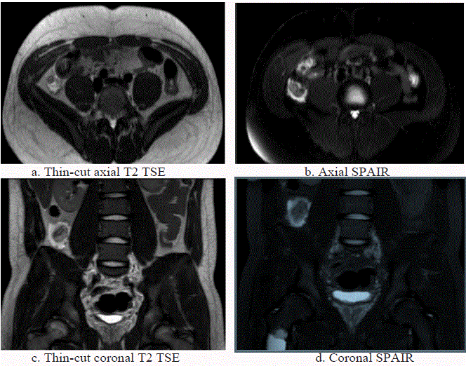
Figure 1
Figure 1
MRI demonstrates a dilated, fluid-filled appendix with thickened, edematous walls and surrounding periappendiceal inflammation, consistent with appendicitis.
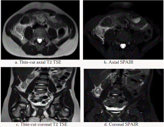
Figure 2
Figure 2
MRI demonstrates a dilated, fluid-filled appendix with thickened, edematous walls and surrounding periappendiceal inflammation. There is additional T2/
SPAIR hyperintensity in the pelvis, consistent with free fluid from perforated appendicitis.
Materials and Methods
This retrospective review of prospectively-collected data is approved by the institutional review board. Pediatric subjects (under the age of 18 years) who presented to the pediatric emergency department from May 23, 2014 to May 22, 2015 with suspected acute appendicitis and who underwent imaging are included in our study. No implemented clinical algorithm is available at our institution to guide imaging selection. The pediatric emergency department physicians determine the imaging modality for each subject. Sixty subjects (17%) initially receive US imaging, while 295 subjects (83%) directly receive cross-sectional imaging (either MRI or CT) without prior US imaging. Three US subjects are diagnosed with acute appendicitis on US, but receive subsequent cross-sectional imaging for treatment planning; these three subjects are excluded from this study. The remaining 57 US subjects have equivocal initial US findings and undergo further imaging (either MRI or CT). These 57 US subjects, as well as the 295 subjects who directly underwent cross-sectional imaging, are included in our study for a total of 352 subjects: 155 subjects who underwent MRI (55% female, mean age of 13 years, age range of 6 to 18 years, 15% with initial US imaging) and 197 subjects who underwent CT (40% female, mean age of 11 years, age range of 2 to 18 years, 17% with initial US imaging).
The decision between MRI and CT for second-line cross-sectional imaging is made by the pediatric emergency department physicians prior to surgical consultation, primarily based on the availability of a 3T MRI system and the subject’s ability to cooperate with breath-hold MRI sequences. A guideline minimum age of 8 years for MRI is used, but younger subjects are selected if deemed capable. Pregnant subjects are scanned on a 1.5T MRI system and are excluded from this study.
All MRI examinations are performed on a 3T MRI system (Ingenia, Philips). Our MRI protocol (Table 1) consists of four breath-hold sequences without intravenous contrast: thin-cut (3-mm slice thickness) axial T2-weighted turbo spin echo (TSE), thin-cut coronal T2 TSE, axial spectral attenuated inversion recovery (SPAIR), and coronal SPAIR. Field of view spans from the right renal upper pole superiorly to the pubic symphysis inferiorly.
All CT examinations are performed on a 256-slice CT (Brilliance ICT, Philips) with intravenous contrast; no oral or rectal contrast is administered. Field of view spans from the dome of the diaphragm superiorly to the pubic symphysis inferiorly.
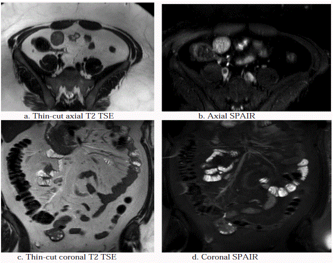
For each subject, radiologic diagnosis and clinical diagnosis are recorded. Age, gender, and department time are also noted. Department time measures the total time spent in the department of radiology by the subject. Sensitivity, specificity, appendix visualization rate, positive appendicitis rate, and alternative diagnosis rate are determined for MRI and CT. Radiologic (for both MRI and CT) diagnosis is graded as “appendicitis” (Figures 1 and 2), “negative” (Figure 3), or “alternative diagnosis.” Imaging findings of appendicitis include dilated appendix (>7 mm in diameter), appendiceal wall thickening (>3 mm), and periappendiceal inflammation/free fluid. Any mention/suggestion of appendicitis (including “possible” or “early” appendicitis) in the impression of the report is considered “positive.” An examination with a non-visualized appendix, but no right lower quadrant inflammation, is considered “negative.” Radiologic diagnosis is based on the final report of the exam (not a second review by the authors). Radiologic diagnosis of “appendicitis” on both MRI and CT is treated with appendectomy or abscess drain placement; subjects with radiologic diagnosis of “negative” on both MRI and CT are either discharged home from the pediatric emergency department or admitted overnight for observation, depending on clinical presentation.
Clinical diagnosis (the reference standard) in subjects who underwent appendectomy is established from the operative and pathology reports. Clinical diagnosis is otherwise established by a 30-day chart review to ensure spontaneous resolution of symptoms without medical or surgical intervention. Our chart review is not able to assess for the return of subjects to different facilities for missed diagnoses, a potential limitation.
Figure 3
Figure 3
MRI demonstrates a normal/“negative” T2/SPAIR hypointense appendix without any T2/SPAIR hyperintensity in the right lower quadrant to suggest an
acute inflammatory process.
Table 1
Table 2
Table 3
Results
Mean age for MRI is 13 years, ranging from 6 to 18 years with a standard deviation of 3.3 years; 11 years for CT, ranging from 2 to 18 years with a standard deviation of 4.5 years (p=0.0004), 55% (85 of 155 subjects) for MRI are female, 40% (79 of 197 subjects) for CT (p=0.003), 15% (23 of 155 subjects) have equivocal initial US imaging prior to MRI, 17% (34 of 197 subjects) prior to CT (p=0.27).
Sensitivity and specificity of MRI are 100% and 98%, 99% and 97% for CT (p=0.61 and 0.53), respectively. Appendix visualization rate is 77% (119 of 155 subjects) for MRI, 90% (178 of 197 subjects) for CT (p=0.0002), positive appendicitis rate is 25% (39 of 155 subjects) for MRI, 34% (66 of 197 subjects) for CT (p=0.175), and alternative diagnosis rate is 3% (5 of 155 subjects) for MRI, 3% (5 of 197 subjects) for CT (p=0.175).
Alternative diagnoses on MRI include one case (0.6%) each of idiopathic hydronephrosis, pyelonephritis, renal abscess, ovarian torsion due to a large paraovarian cyst, and intussusception with a Meckel diverticulum as its lead point. Alternative diagnoses on CT include two cases (1%) of pyelonephritis, and one case (0.5%) each of pancreatitis, small bowel obstruction, and infected urachal diverticulum.
Mean department time for MRI is 28 minutes, ranging from 11 to 48 minutes; 15 minutes for CT, ranging from 3 to 51 minutes (p=0.007).
True positives and true negatives of the 155 subjects imaged with MRI (Table 2), 39 subjects (25%) are correctly diagnosed with acute appendicitis (true positives) and 114 subjects (74%) are correctly diagnosed with no acute appendicitis (true negatives). Of the 197 subjects imaged with CT (Table 3), 66 subjects (34%) are true positives and 126 subjects (64%) are true negatives.
False negative on MRI, no case of acute appendicitis is missed (no false negative). On CT, one subject (a 14-year-old male) is a false negative (0.5%). The initial CT reports a normal appendix. However, due to concerning clinical findings, the subject is admitted for observation. Repeat imaging on hospital day #3 demonstrates perforated appendicitis with a new periappendiceal abscess.
False positives on MRI, two subjects (1%) are diagnosed with early acute appendicitis, but the appendix in both subjects is normal on operative and pathology reports (false positives). On CT, four subjects (2%) are false positives; two subjects resolve clinically with observation only and with no intervention, and two subjects undergo appendectomies that report normal appendix.
Discussion
This study supports 3T MRI as a comparable modality to CT in the evaluation of suspected acute appendicitis in the pediatric population. Although MRI visualizes the appendix at a lower rate than CT (77% for MRI; 90% for CT, p=0.0002), our MRI protocol maintains 100% sensitivity with no false negatives. In fact, the one false negative in this study is a subject who underwent CT. Specificity is similar between the two modalities (98% for MRI, 97% for CT, p=0.53).
Our appendix visualization rate with 3T MRI without intravenous contrast (77%) is an improvement from published data from non-contrast-enhanced protocols on both 1.5T and 3T MRI systems (36% to 43%) [3,4] as well as a contrast-enhanced protocol on a 1.5T system (67%) v[5]. Our mean department time for MRI of 28 minutes is longer than that for CT of 15 minutes (p=0.007) and the published data of 9 to 19 minutes [3-5]. However, the longer department time is clinically insignificant and spares the patient the ionizing radiation and intravenous contrast of CT. Unfortunately, our department times are calculated based on the technologists’ timestamps, which may vary in reliability. Also, our “department time” measures the total time spent in the radiology department by the subject, while published data may measure different time intervals, limiting the ability to compare our data to the published data.
A potential contributing factor to our improved appendix visualization rate when compared to published data is the use of thin-cut T2 TSE sequences. The 3-mm slice thickness is equivalent to that of CT and is less than that of most published MRI protocols [3,5], allowing for improved visualization when compared to published MRI protocols with thicker slices. Our pediatric emergency department physicians are more confident in our radiologic “negative” diagnosis when we visualize a normal appendix. Although our mean examination time is longer to acquire these thin-cut sequences, our improved appendix visualization rate justifies the longer scan time.
Ultimately, the limitations of MRI are scanner availability and subject cooperation. At our institution, we do not have an MRI technologist on site overnight and on weekends. The pediatric emergency department physicians occasionally call in the MRI technologist from home, but typically select CT for subjects during these times. CT technologists are on site at all times; therefore, CT is always available.
The other potential limitation of MRI is the subject’s ability to cooperate. Our MRI protocol consists of breath-hold sequences, which requires cooperation from the subject. Subjects younger than 8 years of age and those in severe pain may not be capable of breath holds, excluding these subjects from MRI. In our study, all of the subjects who begin the MRI complete the examination with satisfactory image quality; no MRI is aborted due to inability to cooperate. Although our protocol is limited in its application in these subjects and increases the examination time, the 100% sensitivity and improved appendix visualization rate support its use.
3T MRI is an effective imaging modality in the evaluation of suspected acute appendicitis in the pediatric population without using ionizing radiation and intravenous contrast. However, prospective research is warranted to confirm our protocol’s accuracy. Due to inherent selection bias of the ordering pediatric emergency department physicians, the demographics of the MRI subjects are statistically older (p=0.0004) and more female (p=0.003) than the CT subjects. The older subject population may be a potential confounding variable, since older subjects may have larger anatomy, enabling easier appendix visualization in our MRI subjects. Understandably, younger subjects are less likely to comply with the breath-hold instructions for MRI and more likely to undergo CT. However, the rates of appendicitis and alternative diagnosis between the MRI and CT subjects are comparable (p=0.175). In addition, a modified MRI protocol for subjects unable to cooperate with breath-hold sequences needs to be developed and assessed. In summary, our study supports 3T MRI as a comparable modality to CT in the evaluation of suspected acute appendicitis in the pediatric population.
References
- Sivit CJ, Applegate KE, Berlin SC, Myers MT, Stallion A, Dudgeon DL, et al. Evaluation of suspected appendicitis in children and young adults: helical CT. Radiology. 2000;216(2):430-3.
- Garcia Peña BM, Mandl KD, Kraus SJ, Fischer AC, Fleisher GR, Lund DP, et al. Ultrasonography and limited computed tomography in the diagnosis and management of appendicitis in children. JAMA. 1999;282(11):1041-6.
- Butler M, Servaes S, Srinivasan A, Edgar JC, Del Pozo G, Darge K, et al. US depiction of the appendix: role of abdominal wall thickness and appendiceal location. Emerg Radiol. 2011;18(6):525-31.
- Doria AS, Moineddin R, Kellenberger CJ, Epelman M, Beyene J, Schuh S, et al. US or CT for diagnosis of appendicitis in children and adults? A meta-analysis. Radiology. 2006;241(1):83-94.
- Brenner DJ, Hall EJ. Computed tomography - an increasing source of radiation exposure. N Engl J Med. 2007;357:2277-84.
- Moore MM, Gustas CN, Choudhary AK, Methratta ST, Hulse MA, Geeting G, et al. MRI for clinically suspected pediatric appendicitis: an implemented program. Pediatr Radiol. 2012;42(9):1056-63.
- Johnson AK, Filippi CG, Andrews T, Higgins T, Tam J, Keating D, et al. Ultrafast 3-T MRI in the evaluation of children with acute lower abdominal pain for the detection of appendicitis. AJR Am J Roentgenol. 2012;198(6):1424-30.
- Duke E, Kalb B, Arif-Tiwari H, Daye ZJ, Gilbertson-Dahdal D, Keim SM, et al. A systematic review and meta-analysis of diagnostic performance of mri for evaluation of acute appendicitis. AJR Am J Roentgenol. 2016;206(3);508-17.
- Koning JL, Naheed JH, Kruk PG. Diagnostic performance of contrast-enhanced MR for acute appendicitis and alternative causes of abdominal pain in children. Pediatr Radiol. 2014;44(8):948-55.
- Hörmann M, Puig S, Prokesch SR, Partik B, Helbich TH. MR imaging of the normal appendix in children. Eur Radiol. 2002;12(9):2313-6. Rosines LA, Chow DS, Lampl BS, Chen S, Gordon S, Mui LW, et al. Value of gadolium-enhanced MRI in detection of acute appendicitis in children and adolescents. AJR Am J Roentgenol. 2014;203(5):543-8.


